FIGURE 6:
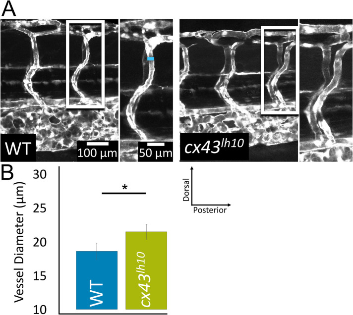
cx43lh10 zebrafish embryos exhibit defects of the vasculature. To evaluate blood vessel morphology in cx43lh10 embryos, cx43lh10 fish were crossed with TG(fli1:EGFP) fish in which all cells of the vasculature are GFP labeled. (A) Representative fluorescence images of GFP-tagged intersegmental vessels of 5 dpf WT/TG(fli1:EGFP) (left) and cx43lh10/TG(fli1:EGFP) mutant (right) embryos. Measurements of vessel diameters is indicated by a blue line in A. (B) Quantification of intersegmental vessels indicates significantly larger vessel diameters in 5 dpf cx43lh10 embryos (n = 5 [WT] and 3 [cx43lh10], p ≤ 0.05; error: SEM).

