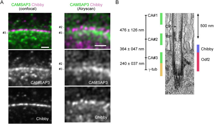
FIGURE 5:
CAMASP3 localization in the TZ. (A) Longitudinal section of a wild-type multiciliated cell, coimmunostained for CAMSAP3 and Chibby, and recorded by confocal or Airyscan microscopy. In these samples, the relative IF intensity of #3 CAMSAP3 puncta is high. Trachea were collected from P180 mice and fixed with methanol. Scale bar, 1 μm. (B) A putative map of CAMSAP3 puncta in relation to the ultrastructure of the axoneme, TZ, and BB. The relative position of each CAMSAP3 punctum was determined by measuring the distances between the puncta, and then the position of the entire CAMSAP3 puncta relative to the ultrastructure was adjusted by placing the #3 punctum at the level of the upper half of Odf2 distribution, as observed in Figure 4. The positions of Chibby, Odf2 and γ-tubulin were estimated referring to previous publications, although γ-tubulin localization needs further confirmation. The scale of the ultrastructure image is shown by a 500 nm bar. The CP is overlaid with a semitransparent white color. CP, the central pair of microtubules; BP, basal plate; TZ, transition zone; TF, transition fiber; BF, basal foot; γ-tub, γ-tubulin; CA, CAMSAP3.

