FIGURE 5:
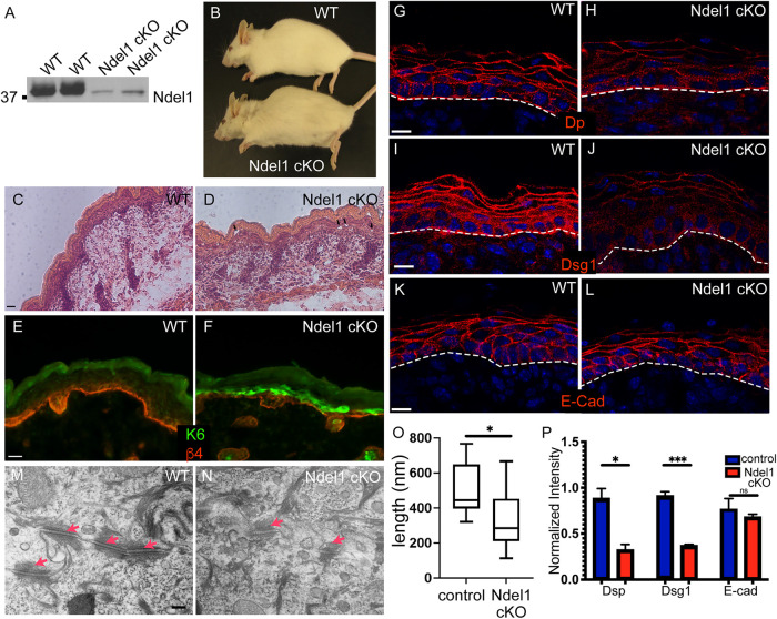
Loss of Ndel1 in the epidermis results in desmosome defects. (A) Western blots of epidermal lysates from two WT and two Ndel1 cKO neonatal mice. (B) Photo of 5-mo-old WT and Ndel1 cKO mice. (C, D) Hematoxylin- and eosin-stained sections from WT and Ndel1 cKO p0 mice. Black arrowheads indicate microblisters in the epidermis. (E, F) Staining for the stress marker keratin 6 (green) and the basement membrane marker β-4 integrin (red) in WT and Ndel1 cKO p0 skin. (G–L) Staining of desmosomal and adherens junction components in WT and Ndel1 cKO p0 skin sections as indicated. All scale bars are 10 μm. (M, N) TEM of spinous cells in control and Ndel1 null epidermis. Red arrows indicate desmosomes. (O) Quantitation of desmosome length in control and Ndel1 null epidermis. n = 25 desmosomes for each, p value is <0.05; Student’s t test. (P) Quantitation of cortical levels of desmoplakin (Dsp), Dsg1, and E-cadherin in control and Ndel1 null epidermis. n = 3, p values are 0.0108 for Dsp, 0.0004 for Dsg1, and 0.534 for E-cad; Student’s t test.

