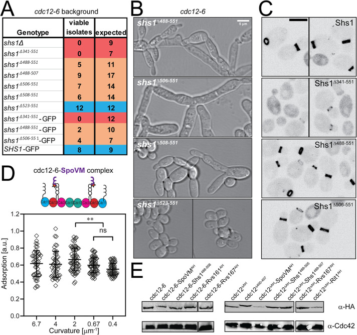
FIGURE 3:
Genetic analyses of Shs1 and Cdc12 AH domains. (A) Viability of cdc12-6 cells expressing indicated SHS1 alleles expressed from the endogenous locus (with 3xHA tag, unless indicated with GFP tag, which lacks 3xHA) based on tetrad dissections at permissive temperature (24°C). Cells from genotypes labeled in blue appeared normal, without obvious septin defects. Genotypes in orange were sick, with partially or fully penetrant septin defects. Genotypes labeled in red were inviable. (B) DIC images of cdc12-6 shs1 mutants (see A) at 24°C. Scale bar, 5 μm. (C) Heterozygous diploids expressing the indicated Shs1 protein fused to GFP from the SHS1 locus. Scale bar, 5 μm. (D) Scatter plot quantifying cdc12-6-SpoVM septin complex adsorption onto different membrane curvatures. Black bars represent the mean. Error bars are the SD for more than 30 measured beads at each curvature across three replicates. Adsorption of cdc12-6-SpoVM complexes was significantly greater on a membrane curvature of 2 μm−1 than on curvatures of 0.67 and 0.4 μm−1; ** (p < 0.01 and p < 0.0001, respectively). ns, adsorption was not significantly different. (E) Western blot comparing expression of indicated Cdc12 chimeras fused to 3xHA epitope in heterozygous diploids.

