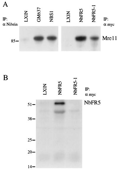
FIG. 3.
Interaction of NbFR5 and NbFR5-1 with Mre11 by immunoprecipitation and Western blot analysis. (A) Total cell lysates were prepared from normal control cell line GM637 and the NBS-ILB1 cell line separately infected with retrovirus carrying the pLXIN vector alone (LXIN), the NBS1 gene (NBS1), the NbFR5 fragment (NbFR5), or the NbFR5-1 fragment (NbFR5-1). Cell lysates were immunoprecipitated (IP) with an antibody directed against nibrin (α Nibrin) or the Myc epitope tag (α myc) and fractionated on a discontinuous polyacrylamide gel. After electrophoretic transfer to a membrane, the blot was probed with a monoclonal antibody to Mre11. The migration of molecular weight markers is indicated on the left (in kilodaltons). (B) Cell lysates from panel A that were immunoprecipitated with an antibody directed against the Myc epitope tag were fractionated on a protein gel and probed with biotinylated anti-Myc antibody.

