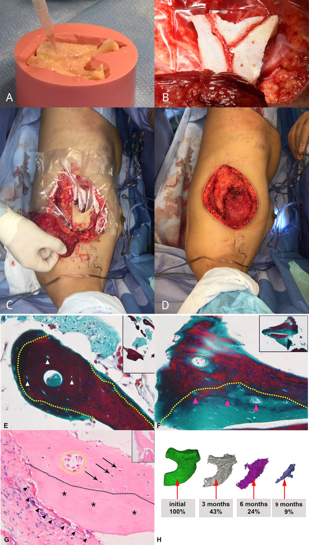
Figure 2.
Graft prefabrication and histological analysis (bone biopsy at moment of transfer at week 32) and volume assessment over time. (A) 3D scaffold of devitalized bone manufactured to match the patient’s defect size and shape and seeded with SVF cells and BMP-2. (B) Ectopic implantation with serratus AV bundle. (C, D) Construct with vascular pedicle before and after being wrapped in split latissimus muscle. (E, F) High magnification of representative figures of bone biopsy after staining with Masson Trichrome. The Tutoplast® scaffold is characterized by purple staining, representing mature bone and cellular lacunae (white arrowheads), showing devitalized bone tissue. Newly formed bone tissue, represented by light green color, is deposited on the Tutoplast® scaffold and contains nuclei (pink arrowheads). The yellow dashed line delineates the original scaffold material and apposition of newly formed bone. A vessel (red circle) demonstrates that the scaffold is vascularized. (G) Overview figure shows appositional bone growth on the Tutoplast® scaffold (asterisk). Osteocytes (arrows) are visible in the newly formed bone. The proportion of scaffold vs. new bone formation is close to 50:50. A blood vessel is present within the newly formed bone (yellow circle). Osteoclasts (full arrowheads) fringe the Tutoplast® scaffold (asterisks), which shows clear signs of degradation at site of interaction. There is no major osteoclast infiltration at the level of the newly formed apposed bone and no sign of degradation visible. (H) CT-reconstruction and volume calculation show volume decrease over time.

