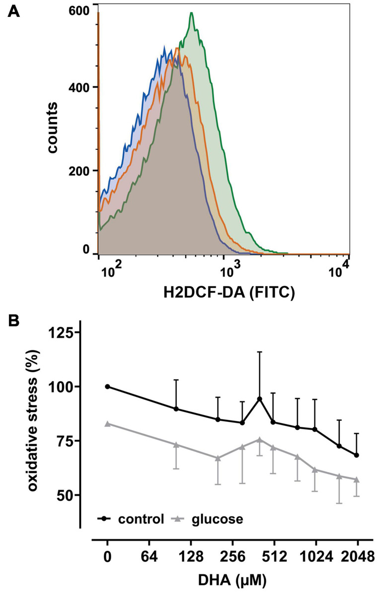
FIGURE 2.

DHA and glucose lower intracellular levels of reactive oxygen species (ROS). (A) Histogram depicting quantitative changes of ROS in erythrocytes treated with 2 mM DHA in the presence (blue) or absence (orange) of 5 mM glucose or PBS only (green). (B) Erythrocytes (n = 6) loaded with 2′,7′-dichlorodihydrofluorescein-diacetate (H2DCF-DA) were treated with various amounts of DHA for 15 min in the presence (gray) or absence (black) of 5 mM glucose at RT and processed for flow cytometry. Mean fluorescence intensity (MFI), reflecting intracellular ROS, is given in percent after normalization to the MFI of PBS control cells (set to 100%).
