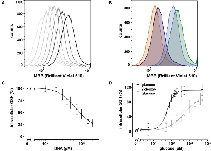
FIGURE 3.
DHA-dependent depletion of cellular glutathione (GSH) is rescued by glucose and 2 deoxy-glucose (2-DG). (A) Histogram of intracellular GSH after incubation with DHA (0.3–2.0 mM, dark to light gray). The black line indicates PBS control cells. (B) Histogram of dose dependent rescue of intracellular GSH upon incubation with 2 mM DHA (orange) in the presence of 100 μM glucose (blue) or 100 μM 2-DG (violet) versus PBS control cells (green). (C) Erythrocytes (n = 11) were treated with various amounts of DHA for 15 min. Cells were then washed and incubated with the fluorescent thiol reagent monobromobimane (MBB) for 10 min. Afterward cells were centrifuged, resuspended in PBS, and processed for flow cytometry. Cells treated for 30 min with 3 mM 1-chloro-2,4-dinitrobenzene (CDNB), a glutathione-S-transferase ρ substrate capable of depleting 96% of cellular GSH within 30 min, were used as negative, cells in PBS as positive controls. MFI values of DHA-treated samples are given as percentages. Data were normalized to the MFI of positive (100%) and negative controls (0%), respectively. (D) Erythrocytes (n = 11) were treated with 2 mM DHA in the presence of indicated concentrations of glucose or 2-DG for 15 min at RT, diluted, and incubated with the fluorescent thiol reagent MBB for 10 min. Afterward, cells were centrifuged, resuspended in PBS, and analyzed by flow cytometry. MFI values are given as percentages after normalizing the data to the MFI of cells pretreated with PBS (100%) or 2 mM DHA without glucose or 2-DG (0%), respectively. Mind that cells treated with 2 mM DHA in the presence of >125 μM glucose have higher intracellular GSH levels than cells treated with PBS only.

