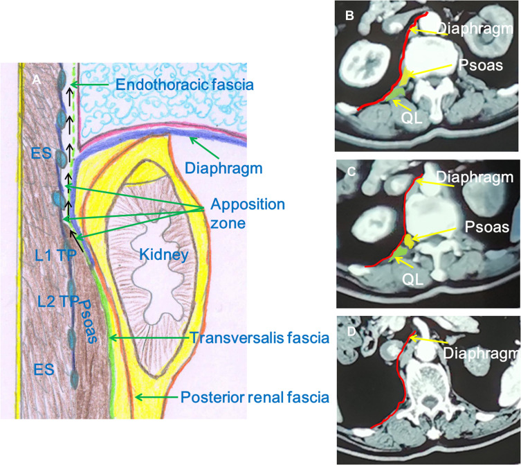
Figure 8.
The communication relationship between the apposition zone of the diaphragm and lower thoracic paravertebral space. (A) The sagittal image at the level of midpoint of L1 TP. The psoas fascia is in continuity with the endothoracic fascia (dashed green line) at the medial arcuate ligament of the diaphragm. The black arrow shows the LA spread from the apposition zone to the lower thoracic paravertebral space. (B–D) The computed tomography shows the cross-sections at the levels of lower edge of L1 vertebral body, upper edge of L1 vertebral body, and T12 vertebral body, respectively. These images show that the apposition zone of the diaphragm at the level of L1 vertebral body gradually develops into the lower thoracic paravertebral space.
Abbreviations: TP, transverse process; LA, local anesthetic; ES, erector spinae; QL, quadratus lumborum.

