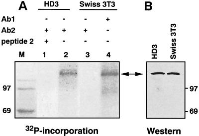
FIG. 2.
CTCF is phosphorylated in vivo. (A) Exponentially growing chicken HD3 and mouse Swiss 3T3 cells were metabolically labeled with [32P]orthophosphate, and CTCF-containing material was immunoprecipitated from cell extracts with 20 μl of protein A-Sepharose 4B-Fast and affinity-purified rabbit polyclonal antibodies (Ab2) specific for the chicken CTCF peptide 2 (lanes 1 to 3) or with pan-specific Ab1 antibodies that recognize CTCF in all vertebrates (26) (lane 4). The Ab2 antibody-blocking peptide 2 (26) was included as the specificity control with chicken cell extracts (lane 1). Immobilized proteins resolved by SDS-PAGE were transferred to membranes and exposed to an X-ray film. (B) The same membranes as in panel A were probed with Ab1 antibodies by ECL immunoblotting assays. The two-headed arrow indicates the position of CTCF. It migrates aberrantly in SDS gels as the 130- to 160-kDa CTCF band (see reference 27 and Results for more details). Lane M, 14C-labeled molecular mass markers.

