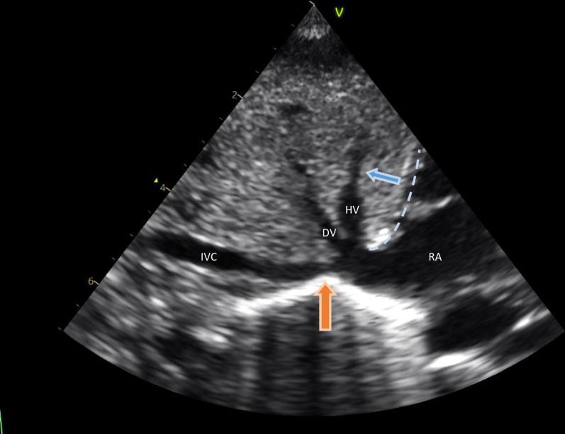
Figure 1.
Subcostal view of the inferior vena cava (IVC) entering the right atrium (RA) with both the ductus venosus (DV) and the hepatic vein (HV) visible. This subcostal view represents the anatomical composition as was observed in all infants. Dashed line: location of the diaphragm. Blue arrow: direction of diaphragm movement with inspiration. Orange arrow: location of IVC collapse, directly caudal to the DV inlet.

