Figure 3.

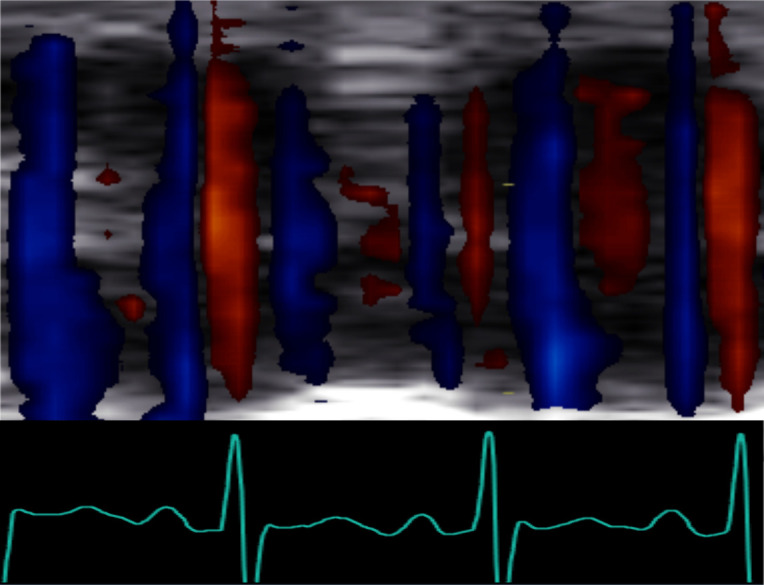
Cross section of the hepatic vein over time using colour Doppler M-mode. Blood flow direction alternates between antegrade (blue) and retrograde (red) with a high frequency and corresponding ECG signal.

Cross section of the hepatic vein over time using colour Doppler M-mode. Blood flow direction alternates between antegrade (blue) and retrograde (red) with a high frequency and corresponding ECG signal.