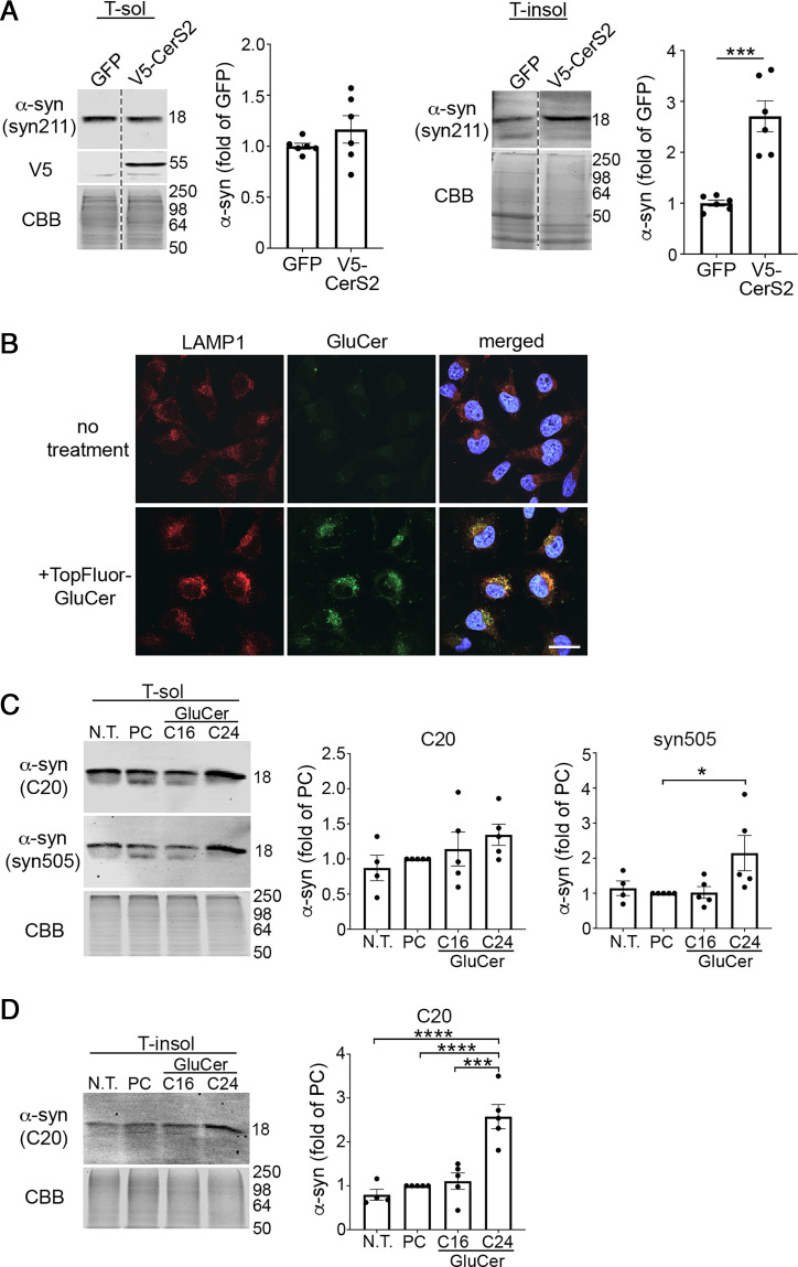
Fig. 5.
Long-chain GSLs induce α-syn pathology in a human cell line and iPSC-derived midbrain neurons. (A) H4 cells were transfected with V5-tagged CerS2 followed by sequential extraction/Western blot to quantify insoluble α-syn (n = 6). (B) Fluorescent-labeled (TopFluor) GluCer (100 μm) was added to H4 cells that overexpress α-syn and uptake was analyzed by confocal microscopy of fixed cells 1 d later. Localization within lysosomes was assessed by LAMP1 immunofluorescence. Nuclei (DAPI) are shown in blue. (Scale bar, 25 μm.) (C and D) iPSC-derived midbrain cultures from healthy subjects aged to day 170 were cultured with 100 μM C16 or C24 GluCer for 3 d followed by sequential extraction and separated into triton X-100 soluble (T-sol) (C) and insoluble (T-insol) (D) lysates followed by Western blot to quantify aggregated α-syn. In C, C20 (rabbit pAb) and syn505 (mouse mAb) were sequentially probed on the same blot and detected with distinct, fluorescent secondary antibodies. Neurons were either not treated (N.T.) or cultured with equimolar PC as controls (n = 5). Values are the mean ± SEM; Student’s t test was used for B; ANOVA with Tukey’s post hoc test was used for C and D; *P < 0.05, ***P < 0.001, and ****P < 0.0001.

