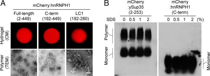
Fig. 1.
In vitro phase separation of hnRNPH1. (A) Hydrogel droplets composed of mCherry-linked full-length (amino acids 2 through 449, Top Left), C-terminal half (C-term, amino acids 192 through 449, Top Middle), or LC1 domain (amino acids 192 through 280, Top Right) of hnRNPH1 were imaged by confocal microscopy (CM) (Materials and Methods). Polymers of each hydrogel droplet sample shown in Bottom panels were visualized by TEM (Materials and Methods). (Scale bar, 0.1 µm) (B) The polymer samples of mCherry-linked ySup35 or hnRNPH1 C terminus were incubated with indicated levels of SDS. The samples were then migrated through the agarose gel containing SDS, and the polymers or monomers were visualized by Western blotting using anti-mCherry antibodies.

