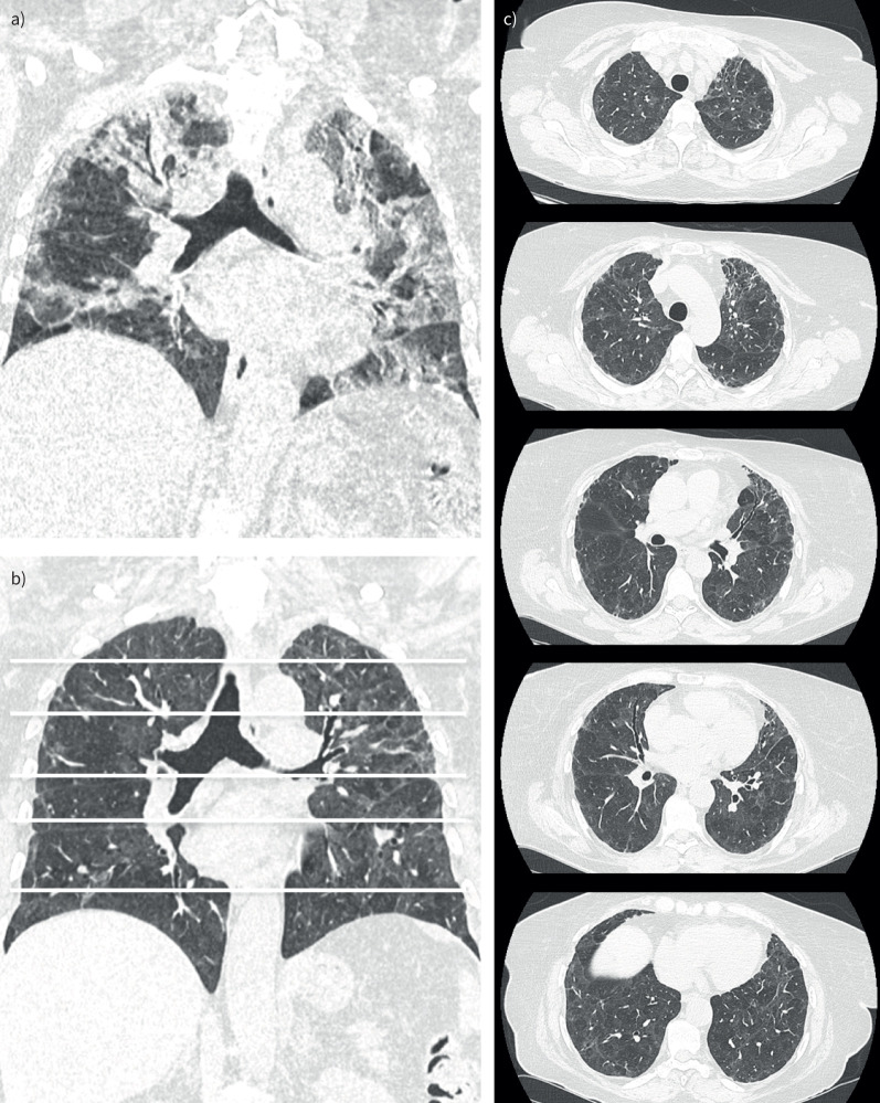
FIGURE 3.
High-resolution computed tomography (HRCT) image of the chest in a patient with severe fibrotic lung lesions 4 months after hospitalisation for coronavirus disease 2019 (COVID-19) compared with that during acute COVID-19. Coronal a) multiplanar reconstruction of an HRCT image of the chest during acute COVID-19 with extensive bilateral ground-glass opacities and consolidations. Coronal b) multiplanar reconstructions and axial sections c) of an HRCT image of the chest from the same patient showing severe fibrotic lung lesions at 4 months, demonstrating diffuse traction bronchiectasis and association with ground-glass opacities.

