. 2021 Dec 2;3(6):e210241. doi: 10.1148/ryct.2021210241

Figure 3:

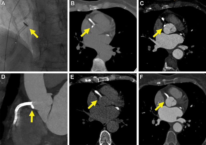
Images in a 72-year-old female patient (patient 3) undergoing intervention for recanalization of a subtotal (99%) in-stent restenosis of the right coronary artery (RCA). There was focal extravasation of contrast media at the RCA ostium (yellow arrow in A). The extravasation was immediately treated with stent extension to the RCA ostium. Thirty minutes after the intervention, CT demonstrated a small, focal, crescent-shaped hyperattenuation of the aortic wall adjacent to the RCA ostium, compatible with subintimal contrast media staining (Dunning I; yellow arrow on B, axial and C, contrast-enhanced CT images and D, multiplanar reformation of contrast-enhanced CT images). Follow-up CT after 24 hours showed complete resolution of the undiluted contrast media accumulation (yellow arrow on E, axial non–contrast-enhanced and F, contrast-enhanced CT images).

Images in a 72-year-old female patient (patient 3) undergoing intervention for recanalization of a subtotal (99%) in-stent restenosis of the right coronary artery (RCA). There was focal extravasation of contrast media at the RCA ostium (yellow arrow in A). The extravasation was immediately treated with stent extension to the RCA ostium. Thirty minutes after the intervention, CT demonstrated a small, focal, crescent-shaped hyperattenuation of the aortic wall adjacent to the RCA ostium, compatible with subintimal contrast media staining (Dunning I; yellow arrow on B, axial and C, contrast-enhanced CT images and D, multiplanar reformation of contrast-enhanced CT images). Follow-up CT after 24 hours showed complete resolution of the undiluted contrast media accumulation (yellow arrow on E, axial non–contrast-enhanced and F, contrast-enhanced CT images).