. 2021 Dec 2;3(6):e210241. doi: 10.1148/ryct.2021210241

Figure 4:

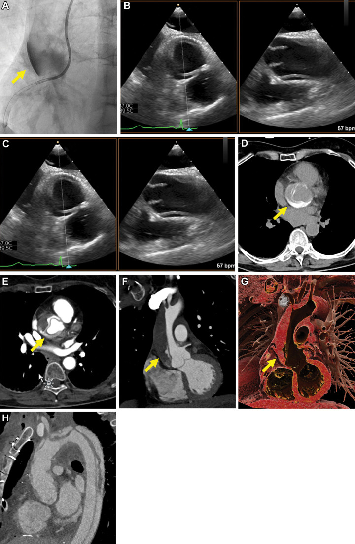
Images in a 70-year-old female patient (patient 4) undergoing recanalization of an acute-on-chronic occlusion of the right coronary artery (RCA). Following balloon predilation of the proximal to mid RCA, extravasation of contrast media in the wall of the ascending aorta occurred (yellow arrow in A). (B, C) Emergency transthoracic echocardiography showed dissection of the ascending aorta. CT performed 30 minutes after percutaneous coronary intervention helped confirm a Stanford type A acute aortic dissection, extending to the origin of an arteria lusoria (Dunning III), with partial layering of hyperattenuated contrast media from the intervention in the aortic root (yellow arrow in D–G). (H) Follow-up CT performed 12 hours after the intervention and after emergency replacement of the ascending aorta and aortic arch showed progression of the dissection to the descending aorta.

Images in a 70-year-old female patient (patient 4) undergoing recanalization of an acute-on-chronic occlusion of the right coronary artery (RCA). Following balloon predilation of the proximal to mid RCA, extravasation of contrast media in the wall of the ascending aorta occurred (yellow arrow in A). (B, C) Emergency transthoracic echocardiography showed dissection of the ascending aorta. CT performed 30 minutes after percutaneous coronary intervention helped confirm a Stanford type A acute aortic dissection, extending to the origin of an arteria lusoria (Dunning III), with partial layering of hyperattenuated contrast media from the intervention in the aortic root (yellow arrow in D–G). (H) Follow-up CT performed 12 hours after the intervention and after emergency replacement of the ascending aorta and aortic arch showed progression of the dissection to the descending aorta.