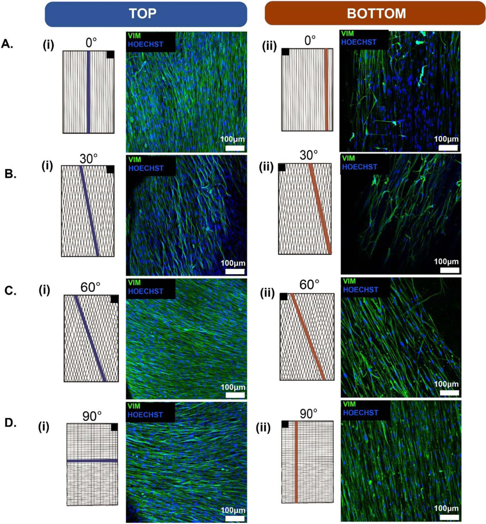
Figure 9.

Immunofluorescence of the composite engineered NHDFs tissues for different PCL fiber architectures. (A) 0° architectured PCL scaffold top and bottom; (B) 30° architectured PCL scaffold top and bottom; (C) 60° architectured PCL scaffold top and bottom; and (D) 90° architectured PCL scaffold top and bottom. In order to image both the front and back, the engineered tissue was flipped along its longitudinal axis. The black box in the upper right (i) or upper left (ii) corner of the schematics are intended to illustrate the direction the engineered tissue was flipped for top and bottom face imaging. Scale bars = 100 μm.
