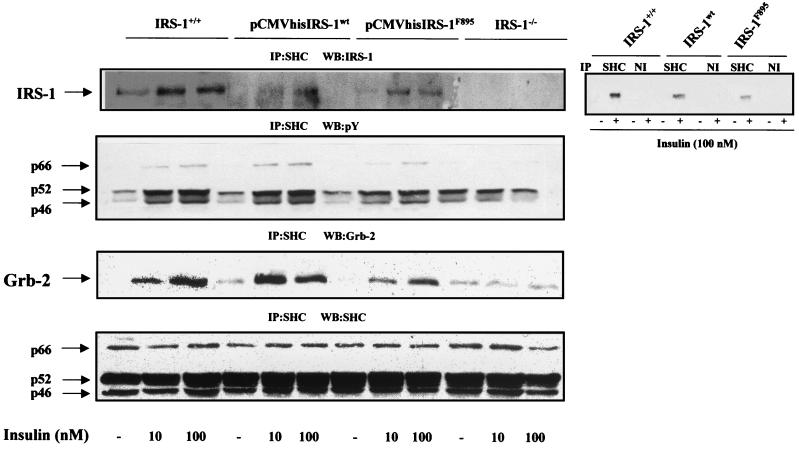
FIG. 7.
Reconstitution of IRS-1wt or IRS-1F895 restored IRS-1–SHC coimmunoprecipitation and SHC signaling. Quiescent cells (IRS-1+/+, IRS-1−/−, and pCMVhis IRS-1wt and pCMVhis IRS-1F895 transfectants) were stimulated with insulin (10 to 100 nM) for 5 min. Control cells were cultured in the absence of hormone. Total cell lysates were immunoprecipitated (IP) with anti-SHC antibody and were subsequently analyzed by Western blotting with anti-IRS-1, anti-Tyr(P), anti-Grb-2, and anti-SHC antibodies as indicated on each panel. The results shown are representative of 4 to 5 experiments, which used two clones of reconstituted cells. As a nonimmune control, cell lysates from untreated and insulin (100 nM)-stimulated wild-type, pCMVhis IRS-1wt, and pCMVhis IRS-1F895 brown adipocytes were incubated with anti-SHC antibody or anti-mouse IgGs and analyzed by Western blotting with the anti-IRS-1 antibody (right panel).

