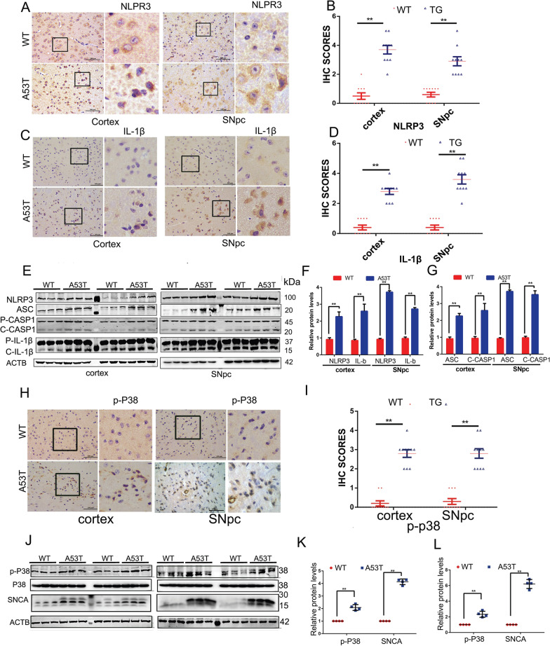
Fig. 1.
NLRP3 inflammasome is activated in the α-synucleinA53T-tg mice. A Immunohistochemistry (IHC) demonstrating increased NLRP3 protein levels in the cortex and SNpc of 9–month-old α-synucleinA53T-tg mice. Scale bars, 100 μm. B Statistical analysis of the scores of NLRP3 staining between α-synucleinA53T-tg and wild-type mice. *p < 0.05 (Student’s t-test). C IHC demonstrating increased IL-1β protein levels in the cortex and SNpc of 9–month-old α-synucleinA53T-tg mice. Scale bars, 100 μm. D Statistical analysis of the scores of IL-1β staining between α-synucleinA53T-tg and wild-type mice. *p < 0.05 (Student’s t-test). E–G Cell lysates from the cortex and SNpc of 9-month-old α-synucleinA53T-tg or wild-type mice were immunoblotted. The protein levels of NLRP3, ASC, cleaved CASP1, IL-1β were statistically analyzed in F and G. Mean ± SEM, n = 6, *p < 0.05 (Student’s t-test). H, I IHC demonstrating increased phosphorylated p38 levels in the cortex and SNpc of 9-month-old α-synucleinA53T-tg mice. Scale bars, 100 μm. I Statistical analysis of the scores of phosphorylated p38 between α-synucleinA53T-tg and wild-type mice. *p < 0.05 (Student’s t-test). J, K Lysates from the cortex and SNpc of mice were immunoblotted using the indicated antibodies. The protein levels of phosphorylated p38 and α-synuclein were statistically analyzed in D and E. Mean ± SEM, n = 6, *p < 0.05 (Student’s t-test)

