Figure 2.

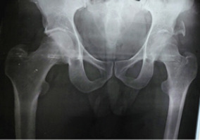
X-ray pelvis anteroposterior postreduction – showing left side posterior wall fracture (Thompson Epstein type 2), concentric reduction seen of both hips.

X-ray pelvis anteroposterior postreduction – showing left side posterior wall fracture (Thompson Epstein type 2), concentric reduction seen of both hips.