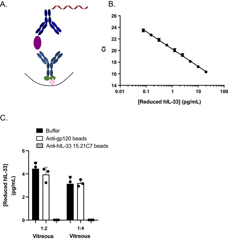
Fig. 3.
Development and specificity of 384-well reduced hIL-33 immuno-PCR (iPCR) assay. A Schematic representation of reduced hIL-33 iPCR assay format: biotinylated rat/human chimeric anti-hIL-33 3F10 capture MAb (light blue), reduced hIL-33 standard (purple), and 55-mer DNA-labeled rat IgG2a anti-hIL-33 15.21C7 detection MAb (dark blue). B Reduced hIL-33 standard curve obtained using iPCR in sample buffer. Data are means ± SD of 6 independent standard curves, each performed with 4 replicates. C Specificity of reduced hIL-33 iPCR assay determined by immunodepletion of reduced hIL-33 in diluted human VH samples using beads conjugated to rat anti-reduced hIL-33 15.21C7 IgG2a (gray bars), control beads conjugated to rat anti-gp120 IgG2a (white bars) and sample buffer (black bars). P = 0.0004 (1:2 VH), P = < 0.0001 (1:4 VH). Data are means ± SD of triplicates

