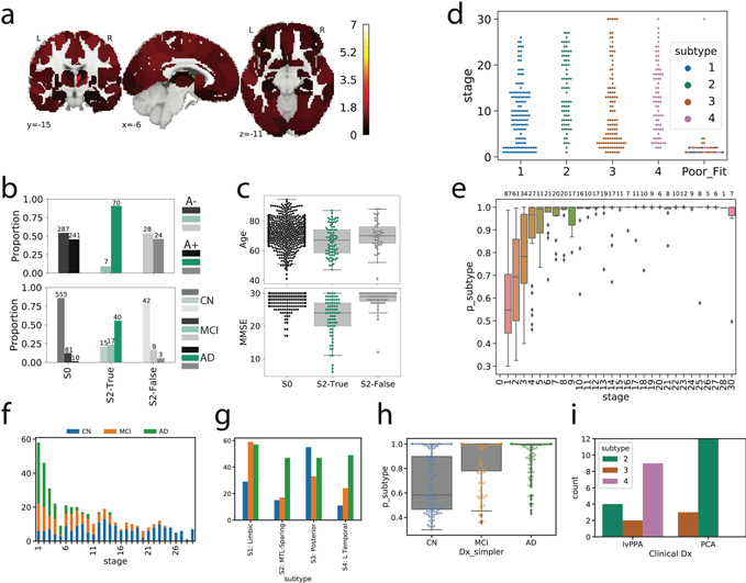
Figure S2.
Details of subtype assignment. a) Several individuals classified as S2 (MTL-Sparing) were found to be tau-negative (i.e. no significant tau in the entorhinal cortex or precuneus). Cortical rendering shows the overall mean tau Z-scores (see Fig S1b) of S2: False individuals. Slightly elevated signal was observed throughout the cortex (but not MTL areas), including in regions where pathological tau is not observed until late AD. b) Proportion of Aβ+ (top) and cognitively impaired (bottom) individuals in S2: False to other S2 individuals (S2: True) and tau-negative individuals (S0). Using, χ2-tests with Tukey’s posthoc correction, a higher proportion of S2: False and S0 individuals were Aβ-and cognitively impaired (ps<0.0001) than S2: True individuals, but did not differ significantly from one another (ps>0.05). c) Using ANOVAs with Tukey’s posthoc correction, S0 and S2: False individuals were older and had higher MMSE scores than S2: True individuals, but did not differ from one another (ps>0.05). d) SuStaIn stage of all individuals stratified by subtype, with the poorly fitting subjects (those that had <0.5 probability of falling into any subtype) shown separately. All but one poorly fit subject exhibited very low SuStaIn stages. e) Probability of maximum likelihood subtype is low at SuStaIn stage 1, but quickly increases with increasing SuStaIn stage. f) Distribution of clinical diagnoses across SuStaIn stages. g) Distribution of clinical diagnoses across subtypes. h) Distribution of maximum-likelihood subtype probabilities for each clinical diagnosis. i) Distribution of PCA and lvPPA subjects from the UCSF sample into each subtype

