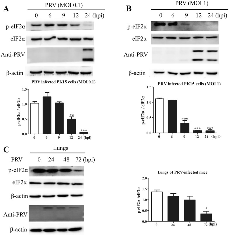
Figure 2.
PRV infection reduced the level of phosphorylated eIF2α in vitro and in vivo. PK15 cells infected with PRV at an A MOI of 0.1 or B MOI of 1.0 were harvested and lysed at 0, 3, 6, 9, 12, and 24 hpi. The levels of p-eIF2α, eIF2α, and PRV proteins were determined by Western blot analysis. The intensities of the p-eIF2α bands were determined by densitometry, normalized to eIF2α, and shown as fold changes (bottom panel). The values are presented as the mean ± SD of triplicate experiments. ** p < 0.01; *** p < 0.001. C BALB/c mice were intraperitoneally inoculated with PRV (1 × 10⁶ PFU) and were then euthanized at 0, 24, 48, and 72 hpi. The levels of p-eIF2α, eIF2α, and PRV proteins in the lungs were determined by Western blot analysis. The intensities of the p-eIF2α bands were determined by densitometry, normalized to eIF2α, and shown as fold changes (right panel). The values are presented as the mean ± SD of triplicate experiments. * p < 0.05.

