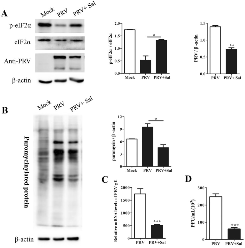
Figure 3.
Salubrinal increased the level of p-eIF2α and suppressed PRV replication in PK15 cells. A PK15 cells were infected with PRV at an MOI of approximately 0.1 in the presence of 100 μM salubrinal. The cells were subjected to puromycin labelling for 1 h at 23 hpi and were then harvested at 24 hpi. Cell lysates were subjected to Western blot analysis to determine the levels of p-eIF2α, eIF2α, and PRV proteins. The intensities of the p-eIF2α bands were determined by densitometry, normalized to eIF2α, and shown as fold changes (right panel). PRV protein expression was quantified by densitometry and normalized to β-actin and are shown as fold changes (right panel). The values are presented as the mean ± SD of triplicate experiments. * p < 0.05; ** p < 0.01. B De novo protein synthesis was measured by using a monoclonal antibody against puromycin; the intensities of bands corresponding to puromycin-labelled proteins were quantified by densitometry and normalized to β-actin and are shown as fold changes (right panel). The values are presented as the mean ± SD of triplicate experiments. * p < 0.05. C, D PK15 cells were infected with PRV at an MOI of approximately 0.1 in the presence of 100 μM salubrinal for 24 h, and supernatants and cells were then harvested. RT–qPCR was performed to determine the relative mRNA level of PRV-gE compared to β-actin (C); the viral titre in the supernatant was determined in PK15 cells (D). The data are presented as the mean ± SD of three independent experiments. *** p < 0.001.

