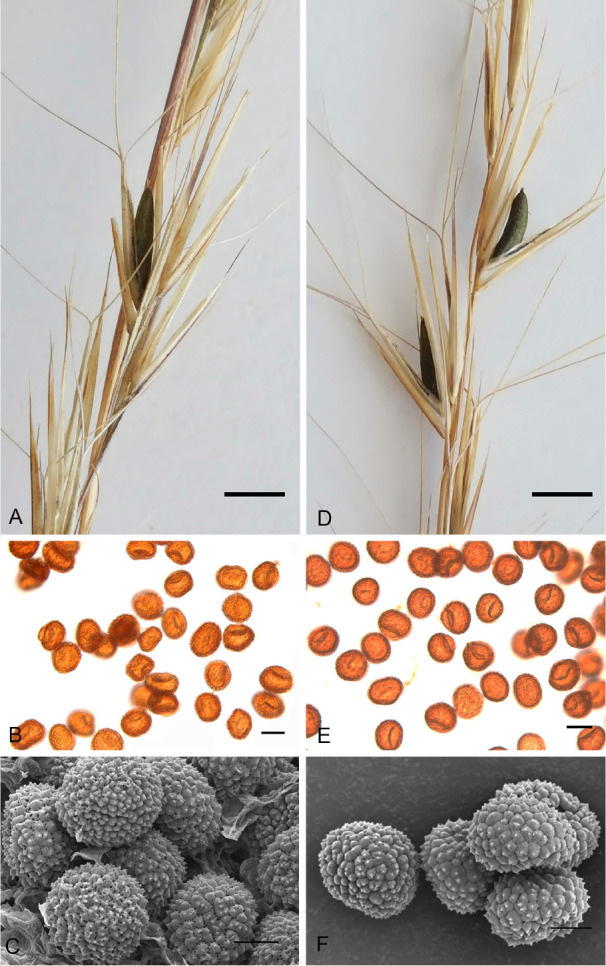
Fig. 3.

Sori and teliospores of Langdonia walkerae sp. nov. on Aristida beyrichiana (A–C) and on Aristida stricta (D–F). A. Sori (WSU 74240). B. Teliospores viewed with transmitted light. C. Scanning electron micrograph (SEM) of teliospores. D. Sori. E. Teliospores viewed with transmitted light. F. SEM of teliospores. Scale bars: A, D = 2 mm; B, E = 10 μm; C, F = 5 μm.
