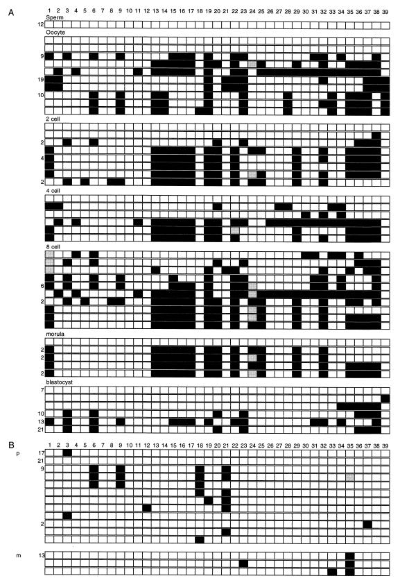
FIG. 5.
Methylation in gametes and embryos. The presentation is the same as in Fig. 3, with the CpG dinucleotides numbered across the top. Paternal (p) and maternal (m) designations are given in the clone sets that came from interspecific crosses. (A) The clones are grouped according to the tissue source. CpG sites 1 to 39 are numbered along the top. (B) Clones from blastocysts derived from (C57BL/6 × M. spretus) crosses.

