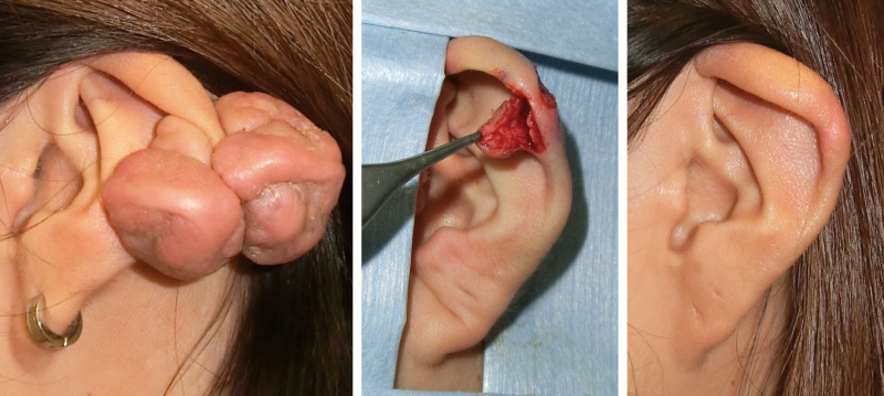
Fig. 8.
Treatment of an auricular keloid by using the core excision method and postoperative radiotherapy. (Left) Preoperative view. (Center) The flap made on the keloid after the core was removed. (Right) Two years after surgery. A 20-year-old woman developed auricular keloids. A flap was designed on the anterior side of the keloid and the core was removed. The flap was sutured using 6-0 polypropylene sutures. After surgery, the site was irradiated (15 Gy, in two fractions, over 2 days). The inflammation dropped uneventfully and the scar became mature over 18 months.

