. 2021 Dec 6;10:e70451. doi: 10.7554/eLife.70451

Figure 1. MEDLE2 is exported to the host cell cytoplasm.

(A) Schematic overview of the chromosomal location for polymorphic gene families in the C. parvum genome. (B) Map of the MEDLE2 locus targeted in C. parvum for insertion of a 3× hemagglutinin (HA) epitope tag, a nanoluciferase reporter gene (Nluc), and neomycin phosphotransferase selection marker (Neo). (C) PCR mapping of the MEDLE2 locus using genomic DNA from wild type (WT) and transgenic (MEDLE2-HA) sporozoites, corresponding primer pairs are shown in (B), and thymidine kinase (TK) gene used as a control. Note the presence of two bands in the 5′–3′ amplification, indicating the presence of a transgene (3081 bp) and persistence of an unmodified copy (1174 bp), suggesting multiple copies of MEDLE2 in the C. parvum genome; also see Figure 1—figure supplement 2. (D, E) HCT-8 cultures were infected with WYLE4-HA (D) or MEDLE2-HA (E) transgenic parasites and fixed after 24 hr for immunofluorescence assay (IFA). Red, antibody to HA; green, Vicia villosa lectin stain, VVL (Gut and Nelson, 1999); blue, Hoechst DNA dye. Additional genes targeted and the localizations of their products are summarized in Table 1 and Figure 1—figure supplements 1 and 3.

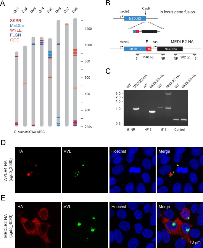
Figure 1.

Figure 1—figure supplement 1. Additional secretory proteins tested in this study.

Figure 1—figure supplement 1.

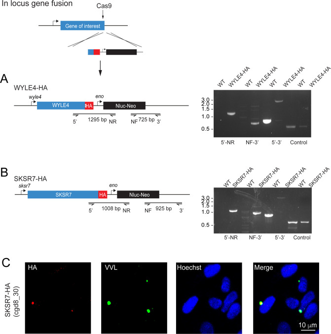
(A–C) Schematic depicting the generation of in-locus gene fusion transgenic parasite strains for WYLE4 (A) and SKSR7 (B). In each case, the C terminus of the gene was targeted for integration of a repair construct containing a hemagglutinin (HA) epitope tag, a nanoluciferase reporter gene (Nluc), and a neomycin phosphotransferase (Neo) selectable marker. Individual primer pairs used to map integration in wild type (WT) and transgenic strains are shown. (C) SKSR7-HA does not localize to the host cell; rather, the protein (red) exhibits expression in the parasite (green).
Figure 1—figure supplement 2. Knockout of MEDLE2 reveals multiple copies of the gene in the genome.

Figure 1—figure supplement 2.

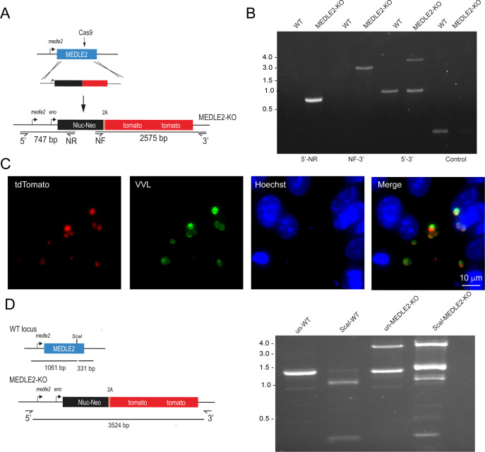
(A) Schematic representation for the strategy used to generate a MEDLE2 KO line, in which the entire locus of MEDLE2 is replaced with a nanoluciferase reporter gene (Nluc) and the neomycin phosphotransferase (Neo) selection marker fused to a 2A peptide, and tdTomato, such that the parasites express a red fluorescent protein in their cytoplasm. The solid black arrow indicates the position of the Cas9-induced double-stranded break in the middle of the gene. Note that this is a different guide from the one used for C-terminal tagging. (B) PCR mapping modification of the MEDLE2 locus using genomic DNA from wild type (WT) and transgenic (MEDLE2 KO) sporozoites using the primer pairs shown in (A) and the thymidine kinase (TK) gene as a control. Note the persistence of a WT band (1392 bp) in the 5′–3′ amplification product, despite the presence of the transgene (3524 bp). (C) HCT-8 cultures were fixed 24 hr after being infected with MEDLE2 KO transgenic parasites. MEDLE2 KO parasites exhibit red fluorescence in their cytoplasm as expected (red, tdTomato, parasite cytoplasm; green, parasites VVL; blue, Hoechst). (D) The full gene PCR products from WT (1392 bp) and MEDLE2 KO parasites (3524 bp) were used for restriction digest with ScaI. A single ScaI restriction site is found in the C terminus of WT MEDLE2; however, integration of the repair cassette disrupts this site. ScaI digested WT PCR product results in two digest products: 331 bp and 1061 bp. Undigested MEDLE2 KO full gene product has the expected 3524 bp fragment, as well as a persisting 1392 bp WT band. ScaI digested MEDLE2 KO shows the 3525 bp repair cassette resistant to ScaI digest, as well as the 331 bp and 1061 bp fragments produced from digest of the unmodified MEDLE2 locus. As a result, there are multiple copies of MEDLE2 in the genome and we have only targeted one for knockout.
Figure 1—figure supplement 3. Other members of the MEDLE gene family are exported to the host cell.

Figure 1—figure supplement 3.

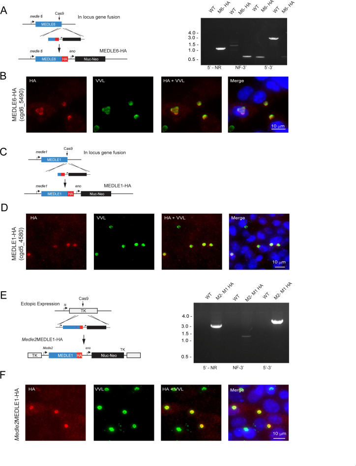
(A) Schematic representation depicting the generation of a MEDLE6-HA transgenic parasite line, in which the endogenous locus of MEDLE6 (cgd6_5490) is a hemagglutinin (HA) epitope tagged at the C terminus. Proper integration at the desired locus was confirmed using PCR mapping with gDNA isolated from MEDLE6 transgenic parasites (M6) and a wild type control (WT). (B) MEDLE6-HA parasites were used to infect HCT-8 cells for an immunofluorescence assay. Cells were fixed every 12 hr and stained for immunofluorescence assay (IFA). Shown as a representative image, at 24 hr post infection, MEDLE6 (red) localizes in/around the parasite (green), as well as slightly in the host cell. Host cell expression is more apparent in multiply infected cells. (C) The MEDLE1 (cgd5_4580) locus was targeted for integration of an HA epitope tag at the C terminus. (D) MEDLE1-HA parasites were used to infect HCT-8 cells for a time-course infection, and IFA was performed on cells fixed every 12 hr. Shown as a representative image, at 12 hr post infection, MEDLE1 (red) localizes in/around the parasite (green), as well as at very low levels in the host cell. (E) Schematic representation for the strategy used to engineer a MEDLE1 overexpression line. The MEDLE2 promoter was used to drive expression of an ectopic copy of MEDLE1-HA expressed in the TK locus. Proper integration was assessed using PCR mapping with gDNA isolated from Medle2MEDLE1-HA (M2- M1 HA) and WT control (WT) parasites. (F) M2-M1 HA parasites were used to infect HCT-8 cells for IFA. At 24 hr post infection, MEDLE1-HA (red) can be seen in/around the parasite (green), as well as in the host cell when expression is driven by the MEDLE2 promoter.