. 2021 Dec 6;10:e70451. doi: 10.7554/eLife.70451

Figure 4. Ordered reporters disrupt MEDLE2 export.

(A) Schematic map of the MEDLE2 locus targeted for insertion of three different reporter genes (mScarlet, beta-lactamase, or Cre recombinase), nanoluciferase (Nluc), and the selection marker (Neo). The guide RNA and flanking sequences used here were the same as those employed to generate MEDLE2-HA transgenic parasites (see Figure 2—figure supplement 1, Figure 4—figure supplement 1 for more detail). (B) MEDLE2-mScarlet parasites were used to infect HCT-8 cells and fixed for immunofluorescence assay (IFA) across a time course. Data shown are from 10 hr post infection, which is representative of the MEDLE2 localization observed at all time points. Red, Medle2-mScarlet; green, parasites (VVL); blue, Hoechst. (C) HCT-8 cells were infected with MEDLE2-BLA C. parvum for 24 hr before incubation with the CCF4-AM beta-lactamase substrate and visualization by live microscopy. This experiment was repeated three times. Red, parasites (tdTomato); green, uncleaved CC4F-AM; blue, cleaved CCF4-AM; gray, DIC. We attribute lack of CCF4-AM cleavage to failure of MEDLE2-BLA to export (Figure 4—figure supplement 1). (D) MEDLE2-Cre parasites were used to infect loxGFP/RFP color switch HCT-8 cells ((Figure 4—figure supplement 2) for schematic representation). After 48 hr, cells were subjected to flow cytometry. Live, single cells were gated based upon forward and side scatter, and green fluorescence (GFP) and red fluorescence (RFP) were measured to detect Cre recombinase activity. Despite robust infection, MEDLE2-Cre-infected cultures did not express RFP (Figure 4—figure supplement 2) compared to the positive control that was transiently transfected to express Cre recombinase.

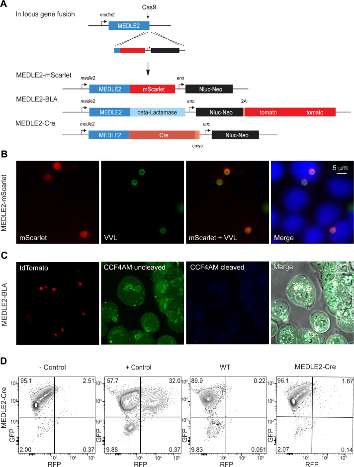
Figure 4.

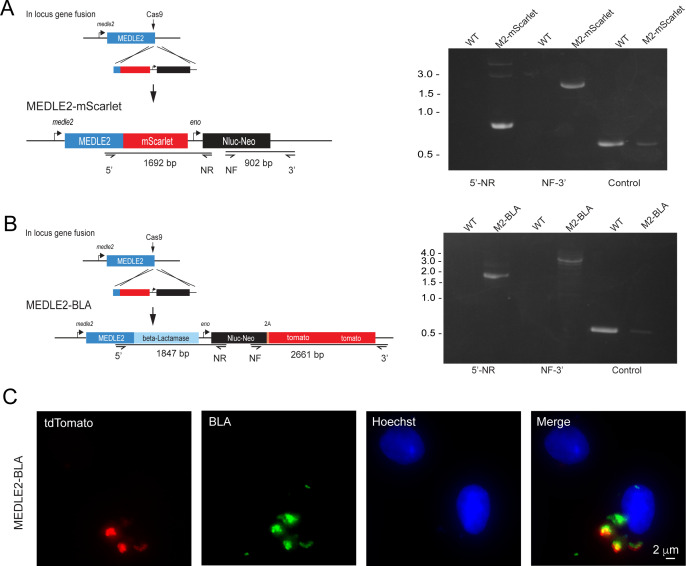
Figure 4—figure supplement 1. MEDLE2-BLA is not exported.

Figure 4—figure supplement 1.

(A) Schematic representation of the strategy used to generate a MEDLE2-mScarlet parasite line, in which the MEDLE2 is C-terminally tagged with the fluorescent protein mScarlet, as well as engineered to express a nanoluciferase reporter gene (Nluc) and the neomycin phosphotransferase (Neo) selection marker. The solid black guide hit sequence is the same as used in Figure 1B to generate the MEDLE2-HA transgenic parasites. Integration PCR using MEDLE2-mScarlet and WT gDNA confirms proper integration. (B) The C terminus of MEDLE2 was targeted for insertion of a construct encoding beta-lactamase (BLA), a nanoluciferase reporter gene (Nluc) and the neomycin phosphotransferase (Neo) selection marker fused to a 2A peptide and a tdTomato reporter gene, using the same tagging strategy previously utilized for this locus. Integration PCR mapping the MEDLE2 locus using genomic DNA from wild type (WT) and transgenic (MEDLE2-BLA) sporozoites with the corresponding primer pairs shown in (A) and the thymidine kinase (TK) gene as a control shows the locus was successfully modified. (C) MEDLE2-BLA transgenic parasites were used to infect HCT-8 cells and fixed at 24 hr for immunofluorescence assay (IFA). Red, parasites (tdTomato); green, beta-lactamase (beta-lactamase antibody); blue, Hoechst.
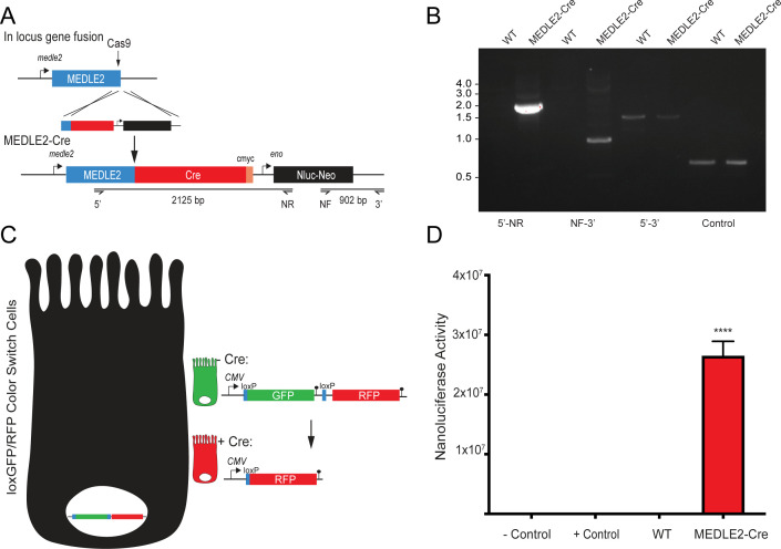
Figure 4—figure supplement 2. MEDLE2-Cre parasites infect loxGFP/RFP color switch cells.

Figure 4—figure supplement 2.

(A) MEDLE2-Cre-expressing parasites were generated by targeting the MEDLE2 locus for insertion of a construct encoding Cre recombinase (Cre), a nanoluciferase reporter gene (Nluc), and the neomycin phosphotransferase (Neo) selection marker. The solid black arrow indicates the guide hit sequence, which is the same guide used in Figure 1B to generate the MEDLE2-HA transgenic parasites. (B) Integration PCR mapping the MEDLE2 locus using genomic DNA from wild type (WT) and transgenic (MEDLE2-Cre) sporozoites using the primer pairs shown in (A) and the thymidine kinase (TK) gene as a control. (C) Schematic of the LoxGFP/RFP color switch HCT-8. Introduction of Cre into these cells induces a color switch from green to red by recombinase-directed removal of the GFP coding sequence along with a stop codon preventing translation of RFP. (D) 1 × 10⁶ transgenic MEDLE2-Cre oocyts were used to infect LoxGFP/RFP color switch HCT-8 cells for 48 hr before preparation of the sample for flow cytometry. 400,000 cells were removed from the sample preparation for nanoluciferase assay to determine infection in the culture. Mean nanoluciferase (relative luminescence) ± SEM is shown for three replicates (p<0.0001; one-way ANOVA with Dunnett’s multiple comparison test). Uninfected cells were used as a negative control, and Cre recombinase transfected cells were used as a positive control.
Figure 4—figure supplement 3. MEDLE2 is an intrinsically disordered protein.

Figure 4—figure supplement 3.

MEDLE2 contains multiple single amino acid repeat regions and lacks a well-defined tertiary structure. The intrinsic disorder of MEDLE2 was assessed using IUPred2A (https://iupred2a.elte.hu/) and the resulting disordered plot is shown as greater than the threshold value of 0.5, represented by the horizontal black line.