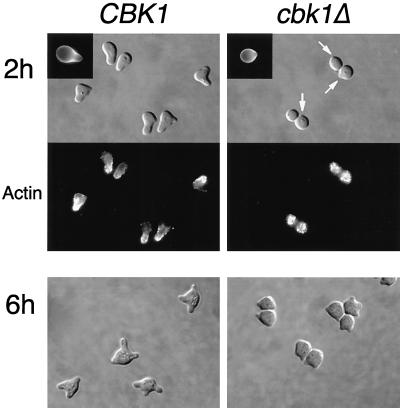
FIG. 6.
Pheromone-induced morphology and actin reorganization in wild-type and cbk1Δ mutant cells. Exponentially growing cells were incubated with α-factor (5 μg/ml) for 2 or 6 h, fixed with formaldehyde, and stained with rhodamine-phalloidin to visualize the actin cytoskeleton. White arrows point out small protrusions on the cell surface. Upper left insets show calcofluor staining of chitin. Strains used for the 6-h time point were bar1Δ mutants. Bar, 5.0 μm.

