
Figure 3.
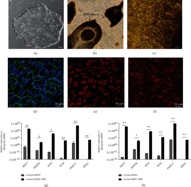
Differentiation of hiPSCs to hiPSC-RPE cells. (a) Representative brightfield image of a parental carrier hiPSC colony, scale bar = 100 μm. (b) Formation of pigmented RPE islands in day 30-40 retinal differentiation cultures, scale bar = 100 μm. (c) Cell culture expansion of RPE islands formed a purified monolayer of hiPSC-RPE cells after 90 days of differentiation, scale bar = 100 μm. (d) hiPSC-RPE cells cultured on coverslips were fixed in 4% paraformaldehyde/PBS and stained with ZO-1 (green) tight junction expression highlighting the hexagonal shape of hiPSC-RPE cells. Nuclei stained with DAPI (blue). (e, f) Immunofluorescence detection of RPE markers CRX and MITF (red) in hiPSC-RPE cells. (g, h) Verification of differentiation of control and carrier (c.93A>G) hiPSCs to RPE cells by RT-qPCR detection of increased relative gene expression of RPE markers (BEST1, MERTK, MITF, PAX6, PMEL17, and RPE65) (unpaired t-test, n = 3 replicates, ∗P < 0.05; ∗∗P < 0.005).