
Figure 4.
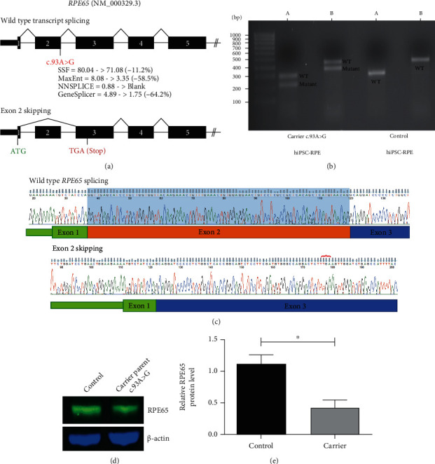
RNA and protein studies of the RPE65 c.93A>G variant. (a) Schematic of wild-type and mutant transcripts and location of the novel c.93A>G variant are shown. Predicted reduction in splice donor strength due to this variant using Alamut Visual is annotated beneath. The site of the predicted premature stop codon (TGA) is indicated in red on the mutant transcript schematic. (b) Representative agarose gel image of cDNA studies illustrating additional presence of a smaller band consistent with exon skipping in c.93A>G carrier hiPSC-RPE cells compared with control. A: amplicon primers located in 5′UTR and exon 4 (mutant allele = 215 bp; wild-type allele = 297 bp); B: amplicon primers located in 5′UTR and exon 5 (mutant allele = 368 bp; wild-type allele = 450 bp). (c) Purified gel band sequencing of the control and mutant cDNA sequence shows excision of exon 2 from the mutant transcript. Red bracket indicates the location of the premature stop codon introduced. Blue shading indicates normal Exon 2 sequence which is absent from the mutant transcript. (d) Western blot analysis showing RPE65 levels in the control [Left] and parental carrier hiPSC-RPE cells [Right]. (e). Density analysis of RPE65 protein bands shows approximately 50% reduction in RPE65 expression in the parental carrier hiPSC-RPE cells compared with the control (unpaired t-test, n = 3 independent experiments, ∗P < 0.05).