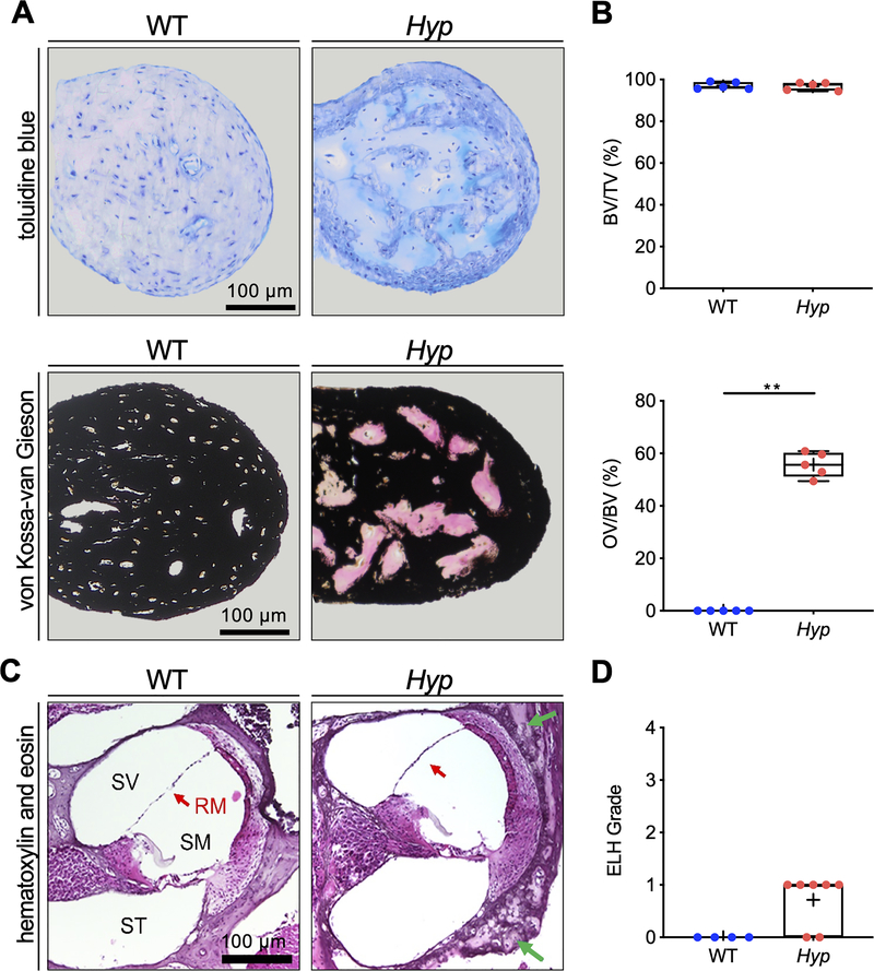
Fig. 4. Undecalcified histology identifies osteoid accumulation in the malleus and signs of mild endolymphatic hydrops in the cochlea in Hyp mice.
(A) Representative histological images of toluidine blue stained orbicular apophysis of the malleus of 24-week-old Hyp and WT mice (upper panel) and von Kossa-van Gieson staining (lower panel). (B) Evaluation of bone volume fraction (BV/TV; upper panel), osteoid volume per bone volume (OV/BV; lower panel) in Hyp mice compared to WT. (C) Representative cochlear cross-sections from 6-week-old male WT (left panel) and Hyp mice (right panel) in hematoxylin and eosin (H&E) staining. The otic capsule in Hyp mice shows osteoidosis (green arrows) and a bulging Reissner’s membrane (RM, red arrow). (D) Endolymphatic hydrops (ELH) in the basal turn classified into the severity grades (23). ELH was not observed in WT animals whereas grade 1 ELH was detected in five out of seven analyzed Hyp animals. **p<0.01 (two-tailed unpaired t-test and Mann-Whitney-U-test).

