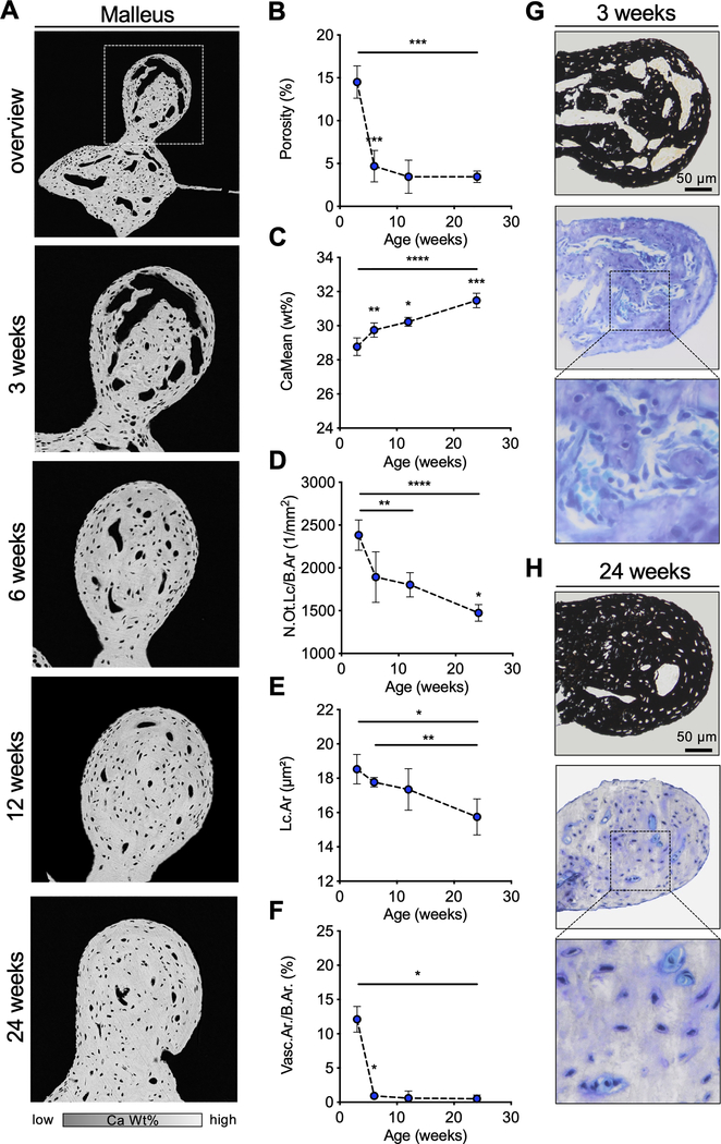
Fig. 5. Postnatal development of the ossicles in WT mice.
(A) qBEI images of the orbicular apophysis of the malleus at selected timepoints. (B, C) Quantification of the osseous porosity, CaMean and CaPeak. (D, E) Analysis of the number of osteocyte lacunae per bone (N.Ot.Lc/B.Ar) and the mean osteocyte lacunar area (Lc.Ar). (F) Quantification of the vascular area per bone area (Vasc.Ar/B.Ar). (G, H) Histological images (von Kossa-van Gieson and toluidine blue staining) of the malleus at the age of 3 weeks and 24 weeks indicating the reduction of porosity with age in WT mice. *p<0.05, **p<0.01, ***p<0.001, ****p<0.0001 (two-tailed paired t-test).

