
Figure 3:
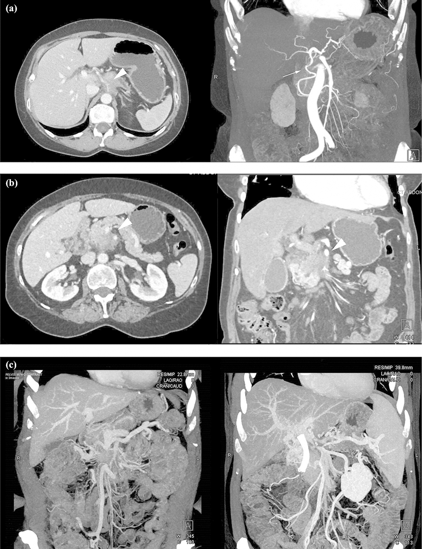
(a) CT imaging of a patient with LAPC-1: encasement of the celiac artery (arrowhead) and no involvement of the gastroduodenal artery (arrow), (b) CT imaging of a patient with LAPC-2: encasement of the SMA and SMV (arrowheads), (c) CT imaging of a patient with PDAC and non-reconstructible SMV, LAPC-3.