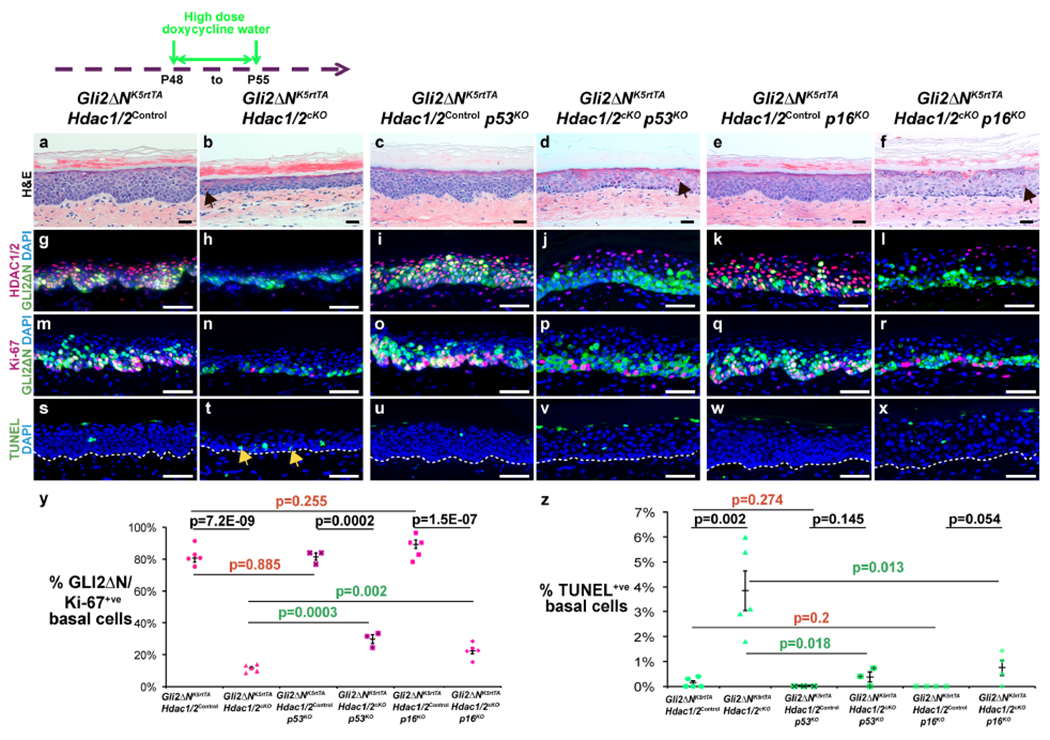
Figure 5 – Hdac1/2 deletion reduces proliferation and increases apoptosis of GLI2ΔN-expressing plantar epidermis via p53- and p16-dependent mechanisms.

Schematic shows timing of doxycycline treatment; samples were analyzed at P55. (a-f) Histology of plantar epidermis for the indicated genotypes. Arrows in (b,d,f) indicate prematurely cornified cells. (g-r) IF for the indicated proteins and genotypes. (s-x) TUNEL staining for the indicated genotypes. Arrows in (t) indicate apoptotic cells. White dashed lines indicate epidermal-dermal boundaries. (y,z) Quantitation of Ki-67-positive (y) or TUNEL-positive (z) GLI2ΔN-expressing plantar epidermal basal cells for the indicated genotypes; n≥3 mice of each genotype were analyzed. P-values calculated with two-tailed Student’s t-test; error bars indicate SEM. Scale bars: 25μm (a-f), 50μm (g-x).
