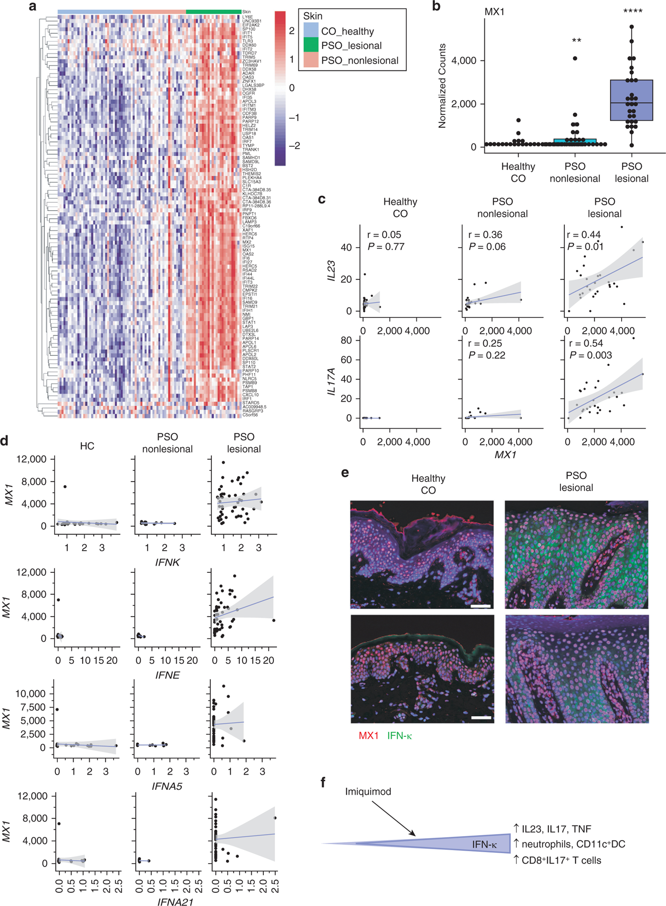
Figure 1. IFN signatures are elevated in PSO skin and correlate with IL23 and IL17A expression.

(a) Heatmap identifying the expression of IFN-1‒regulated genes in healthy CO (left, blue bar), nonlesional PSO skin (middle, pink bar), and lesional PSO skin (right, green bar). (b). Expression of MX1 in the CO, nonlesional, and lesional skin. ** P < 0.01, **** P < 0.0001. (c). Correlations of MX1 with IL23 (top) and IL17 (bottom). Pearson coefficients are shown. (d) Correlations of detectible IFN1 transcripts with MX1 in a second dataset of control and psoriasis biopsies (n = 36 healthy control, 13 nonlesional skin, and 50 lesional skin samples). (e) Immunofluorescent microscopy for MX1 (red) and IFN-κ (green) in two CO and lesional PSO skin biopsies. (f). Graded expression of IFN-κ in the epidermis regulates inflammatory mediators of psoriasis. Increasing baseline IFN-κ results in the upregulation of epidermal proliferation, cellular infiltrates, and IL-17 and IL-23 responses after imiquimod treatment. CO, control; DC, dendritic cell; PSO, psoriatic.
