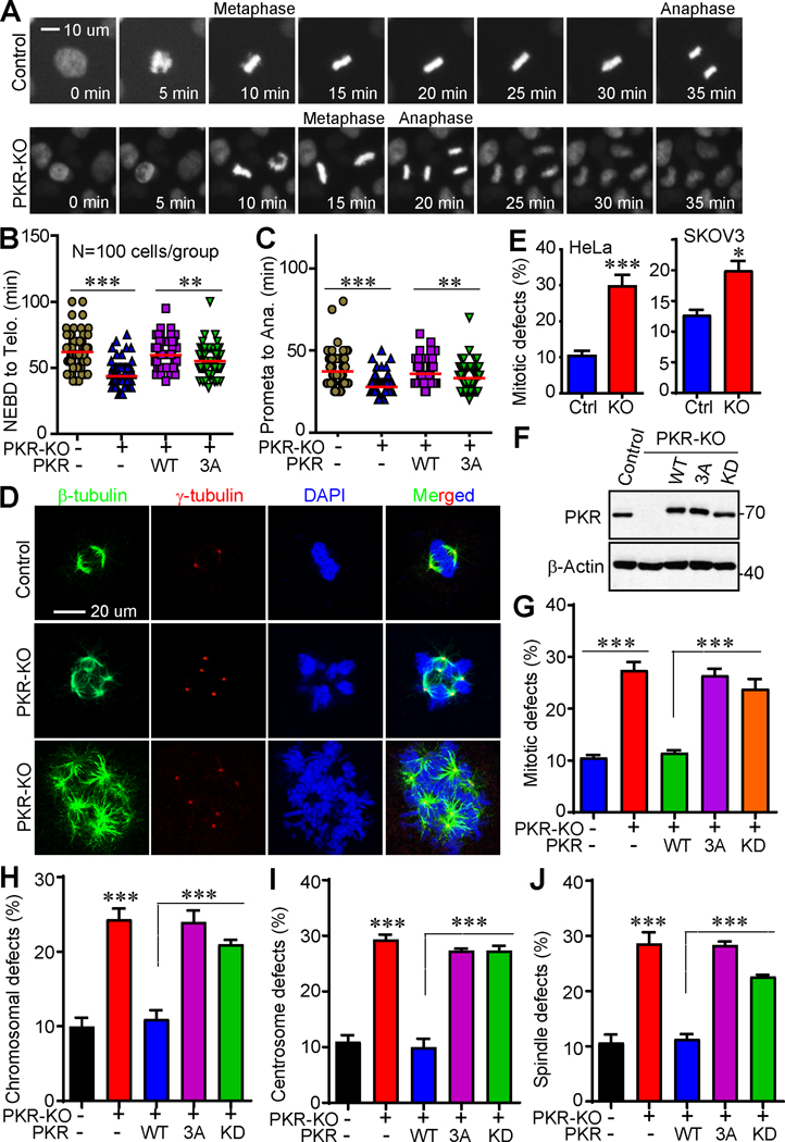
Figure 2. Phosphorylation of PKR is required for precise mitosis in cancer cells.
(A) PKR deletion shortens mitotic length (from NEBD to anaphase onset) in HeLa cells. RFP-HeLa and RFP-HeLa-PKR-KO (knockout) cells were seeded in 96-well plate and incubated overnight. Live-cell imaging was performed using a Fluorescent Microscope Imager with a 20X objective and collected every 5 min for 24 h. Representative cells are shown.
(B, C) Various cell lines were subjected to live-cell imaging and mitotic length was quantified. NEBD: nuclear envelope breakdown; Ana: anaphase onset. Data in B, C were collected from 100 mitotic cells for each group (mean ± SD of three independent experiments). **: p<0.01; ***: p<0.001 (unpaired Student’s t test).
(D, E) Knockdown of PKR resulted in massive mitotic defects in HeLa and SKOV3 cells. Representative photos of normal mitosis (control) and mitotic abnormalities (PKR-KO) in HeLa cells were shown in D. Cells were stained with β-tubulin, γ-tubulin antibodies, and DAPI to visualize microtubules (green), centrosomes (red), and chromosomes (blue), respectively. Data in E were collected from 150 mitotic cells for each group (mean ± SD of three independent experiments).
(F) Establishment of cell lines expressing exogenous PKR-WT, PKR-3A, or PKR-KD (kinase dead) in HeLa PKR-KO cells.
(G-J) Qualification of cells with mitotic defects in cell lines established in F. Data were collected from 150 mitotic cells for each group (mean ± SD of three independent experiments). ***: p<0.001 (unpaired Student’s t test).

