. 2021 Dec 9;63(1):42–55. doi: 10.3349/ymj.2022.63.1.42

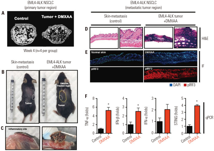
Fig. 1. DMXAA reduced tumor growth and induces strong antitumor response in EML-ALK NSCLC model. (A) MRI images of the primary tumor site of the control and DMXAA (500 µg)-treated tumor-bearing mice. (B) Skin nodule in the EML4-ALK mice. White dotted line indicates the metastatic site, while yellow dotted line indicates the DMXAA-treated region. (C) Skin inflammation observed in the control and DMXAA-treated sites. (D) H&E staining, (E) DAPI (upper) and pIFR3 expression (lower) images of normal skin (control) and DMXAA-treated site. (F) Expression levels of TNF-α, IFN-β, IFN-α, and STING. The results are expressed as mean±SD. *p<0.05. TNF, tumor necrosis factor; IL, interleukin; IFN, interferon; STING, stimulator of interferon genes; H&E, hematoxylin and eosin; qPCR, real-time PCR.

Fig. 1