. 2021 Dec 9;63(1):42–55. doi: 10.3349/ymj.2022.63.1.42

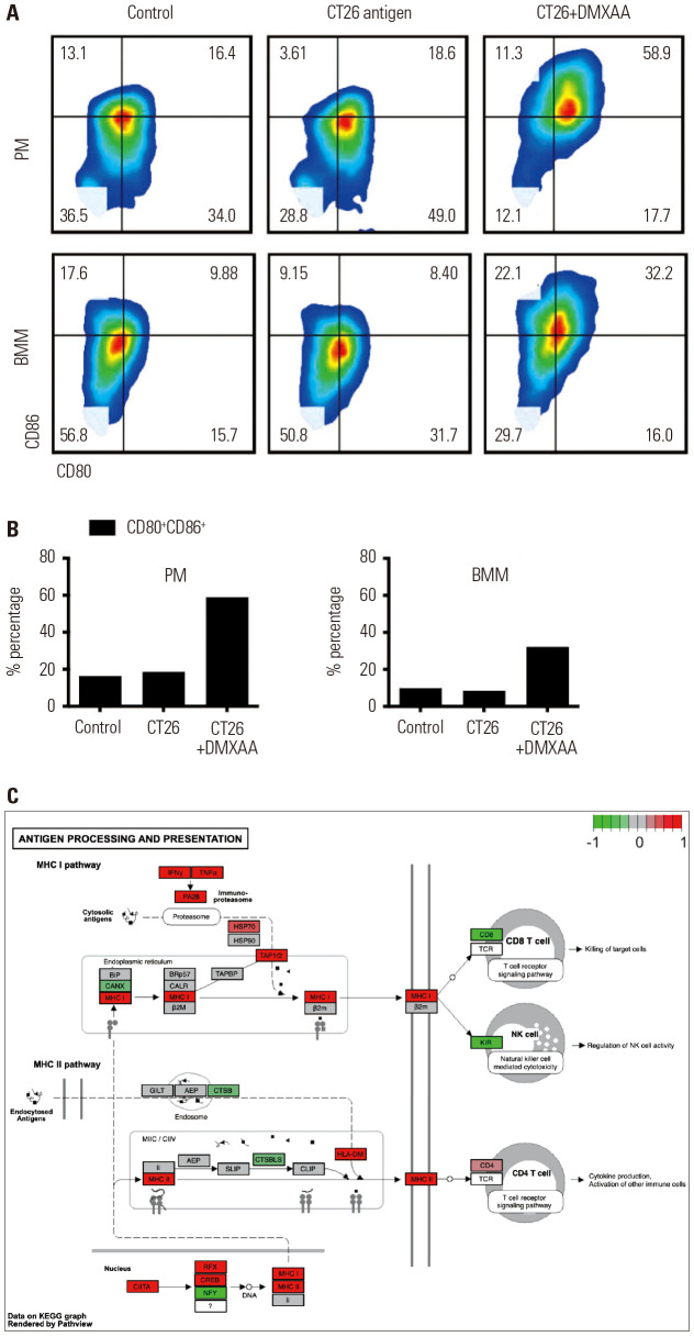
Fig. 4. Reinforcement of tumor antigen-presenting activity via CD80/CD86 after DMXAA treatment. (A) Transient induction of CD80 and CD86 in control (vehicle-treated group), CT26 antigen-treated, and CT26 antigen+DMXAA treated macrophages isolated from the peritoneum and bone marrow. (B) The CD80 and CD86 expression levels on the surface increase in a time-dependent manner. The percentages of CD80 and CD86 double-positive cells increased from 24.4% at 0 h to 66.3% at 24 hours of DMXAA treatment in the PMs (left) and BMMs (right). No changes were observed in the macrophages treated with only a CT26 antigen. (C) DMXAA-related alteration of gene expression associated with antigen processing and presentation in macrophages. The entire MHC I and MHC II signaling pathways are presented. The molecules with higher or lower gene expression compared to the control are highlighted in red and green, respectively. PM, peritoneal macrophages; BMM, bone marrow macrophages; MHC, major histocompatibility complex.

Fig. 4