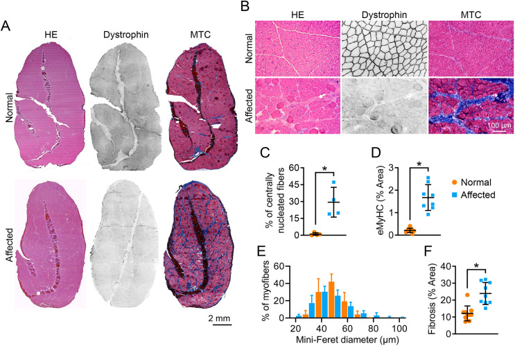
Fig. 2.
Dystrophic ECU muscle showed characteristic muscle pathology. (A) Representative full-view photomicrographs of H&E staining, dystrophin immunostaining and MTC staining from the normal and affected dog ECU muscle. (B) Representative close view photomicrographs of H&E staining, dystrophin immunostaining and MTC staining from normal and affected dog ECU muscle. (C) Quantification of the centrally nucleated myofiber in the normal (n=4) and affected dog (n=4) ECU muscle. (D) Quantification of the eMyHC-stained myofiber area in the normal (n=9) and affected (n=8) dog ECU muscle. (E) Morphometric quantification of the myofiber size in the normal (n=6) and affected (n=7) dog ECU muscle. (F) Percentage of the fibrotic area in the normal (n=10) and affected (n=9) dog ECU muscle. Data are mean±s.d. *P<0.05 (statistical analysis was performed using unpaired Student’s t-test for C, D and F).

