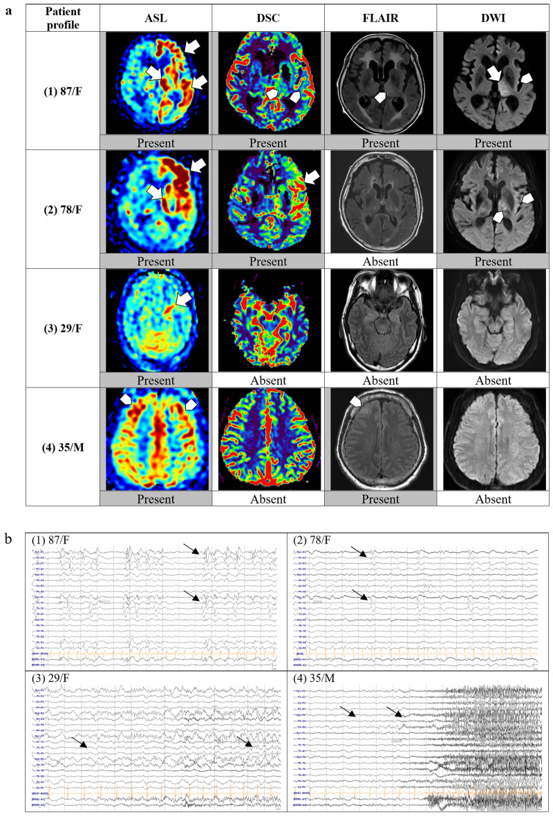
Figure 2.
Examples of brain MRI sequences and EEGs of four representative patients with status epilepticus. Brain MRI and EEG of each patient were listed according to the same number from (1) to (4). (a) Brain MRI: (1) ASL showing hyperperfusion in the left frontal and temporal cortices and the left thalamus. DSC reveals subtle increases in perfusion in the left thalamus and lateral temporal lobe. FLAIR and DWI show changes in the left thalamus and insula. (2) ASL and DSC show definite hyperperfusion in the left frontal and temporal lobes, FLAIR shows no changes, and DWI shows subtle restrictions in the left thalamus and insula. (3) ASL shows definite hyperperfusion in the left hippocampus, but no signal changes are noted on DSC, FLAIR, or DWI. (4) ASL showing subtle hyperperfusion in the bilateral frontal cortices, and subtle T2 hyperintensity is noted in the left frontal cortex. (b) EEG: (1) Left frontotemporal periodic discharges. (2) Left frontotemporal periodic discharges. (3) Left temporal rhythmic delta activity with evolution indicating an electrographic seizure. (4) Right frontal theta rhythm evolving to beta activity, indicating an electrographic seizure. ASL arterial spin labeling; DSC, dynamic susceptibility contrast, DWI diffusion-weighted imaging, EEG electroencephalogram, FLAIR fluid-attenuated inversion recovery, MRI magnetic resonance imaging.

