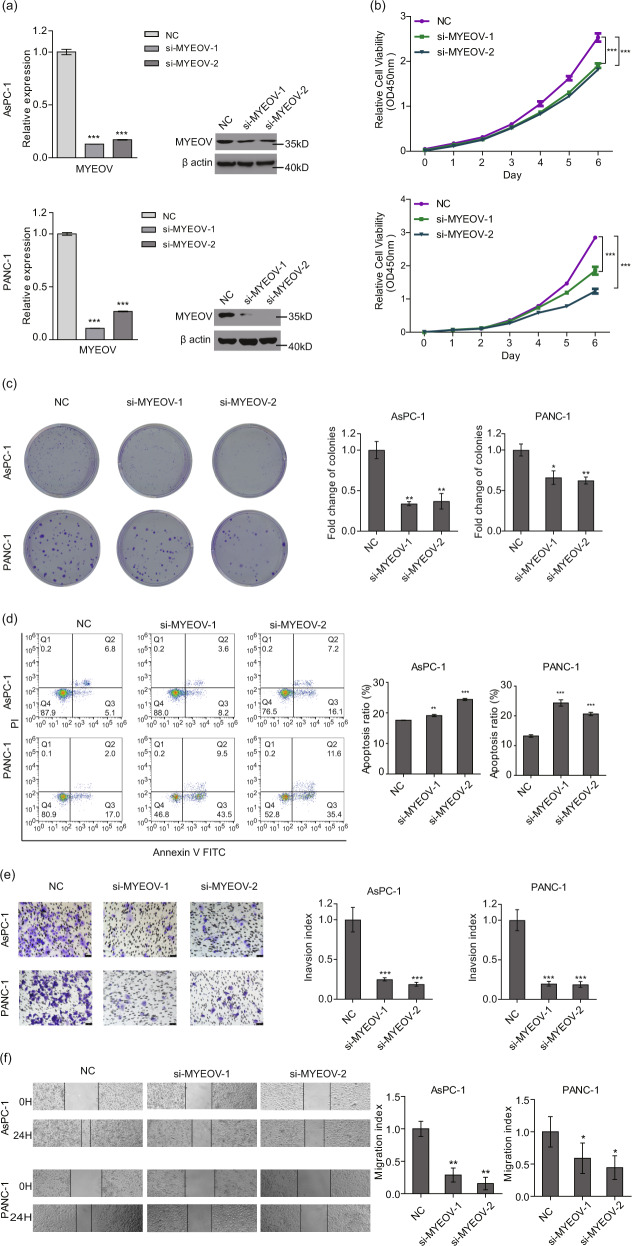
Fig. 2. MYEOV knockdown inhibits cell proliferation, invasion and migration in PDAC in vitro.
a The expression levels of MYEOV were quantified by qPCR and Western blot. b, c Cell proliferation measured by the CCK-8 (b) and colony formation assays (c). d Apoptosis as determined by flow cytometry. e, f Cell invasion (e) and migration (f) as determined by transwell invasion assays (200× magnification) and scratch assays (40× magnification). All n ≥ 3; bar, SEM; *P < 0.05, **P < 0.01, ***P < 0.001 and ****P < 0.0001 compared with NC; Student’s t-test.

