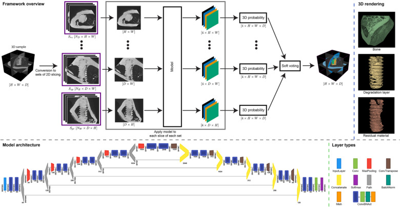
Figure 1.
Overview of the segmentation framework for high-resolution synchrotron radiation microtomograms. Top shows the full segmentation pipeline: 1. conversion of 3D tomograms into 2D slices. 2. processing of slicing sets by our model. 3. soft voting is used to fuse the multi-axes prediction into the final segmentation. Top right: 3D rendering of the resulting segmentation (created with 3D Slicer, v4.11, https://www.slicer.org/). Bottom shows our final U-net model architecture and the layer legend (created with Net2Vis38, https://github.com/viscom-ulm/Net2Vis).

