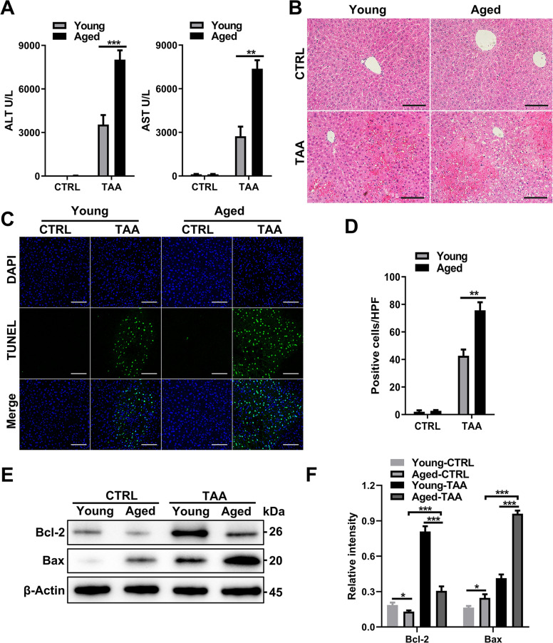
Fig. 1. Aging exacerbates liver injury and inflammation after TAA treatment.
Young and aged mice were administered TAA (200 mg/kg) and PBS as a control. A Serum ALT (left panel) and AST (right panel) levels in each group. B Representative H&E staining of the liver. C Representative TUNEL (green fluorescence) staining of the liver with DAPI counterstaining (blue fluorescence) and D quantification. E Immunoblotting of Bax and Bcl-2 expression in liver tissue from each group and F quantification. All data shown represent n = 8–10 mice per group. All results are representative of at least three independent experiments. Values are presented as the mean ± SD. *p ≤ 0.05; **p ≤ 0.01; ***p ≤ 0.001.

