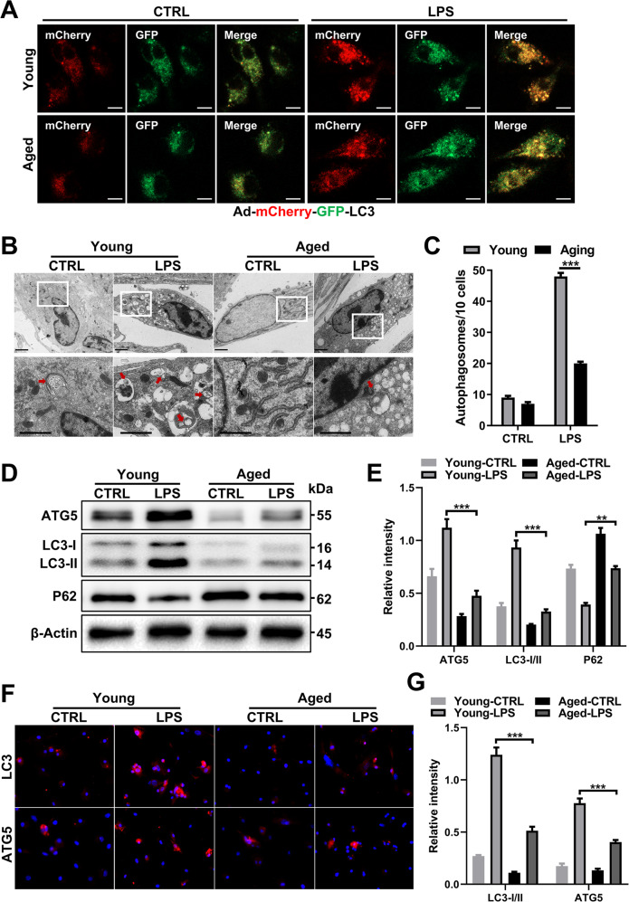
Fig. 3. Aging impairs autophagy in LPS-treated macrophages.
Young and aged BMDMs derived from mouse bone marrow were infected with Ad-mCherry-GFP-LC3 (20 MOI) on the 5th day when the cells were not fully mature and then treated with LPS (100 ng/ml) on the 7th day. A Representative laser confocal microscopy images. B Transmission electron microscopy (TEM) examination of autophagosomes from young and aged BMDMs and (C) quantification. D Immunoblotting of LC3-I/II, ATG5 and p62 expression in BMDMs from each group. Quantification in (E). F Representative IF staining of LC3-I/II and ATG5 incorporation (red fluorescence) with DAPI counterstaining (blue fluorescence) in BMDMs. Quantification in (G). All results are representative of at least three independent experiments. Values are presented as the mean ± SD. *p ≤ 0.05; **p ≤ 0.01; ***p ≤ 0.001.

