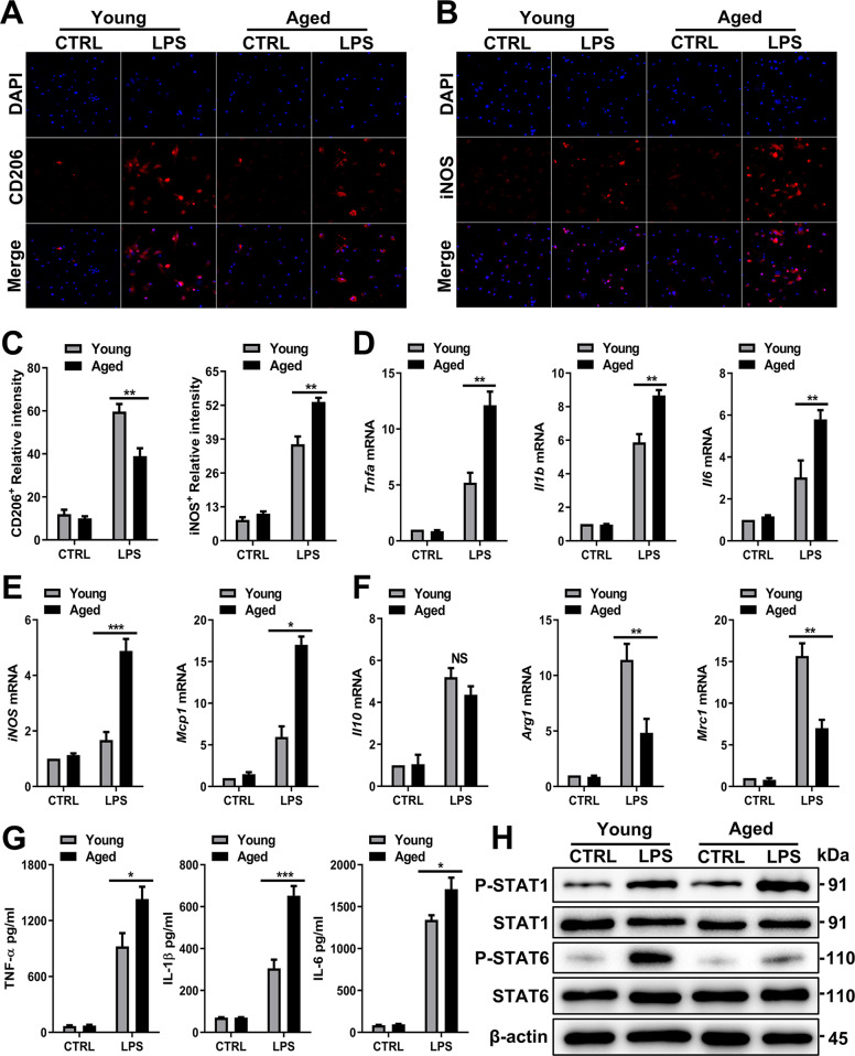
Fig. 5. Aged macrophages preferentially polarize into the M1 phenotype.
Young and aged BMDMs were treated with LPS (100 ng/ml) and PBS as a control. A Representative IF staining of CD206 incorporation (red fluorescence) in the liver with DAPI counterstaining (blue fluorescence). B Representative IF staining of iNOS incorporation (red fluorescence) in the liver with DAPI counterstaining (blue fluorescence). C Quantification of A (left) and B (right). D, E Proinflammatory mRNA expression (Tnfa, Il1b, Il6, iNOS, and Mcp1) and (F) anti-inflammatory mRNA expression (Arg1, Il10, and Mrc1) in BMDMs were detected by RT–PCR. The average target gene/GAPDH ratios of different experimental groups relative to the control group are given. G Determination of the total protein content (TNF-α, IL-1β, and IL-6) in the culture supernatant of BMDMs after LPS treatment by ELISA. H Immunoblotting of STAT1, P-STAT1, STAT6, and P-STAT6 expression in BMDMs from each group. All results are representative of at least three independent experiments. Values are presented as the mean ± SD. *p ≤ 0.05; **p ≤ 0.01; ***p ≤ 0.001.

