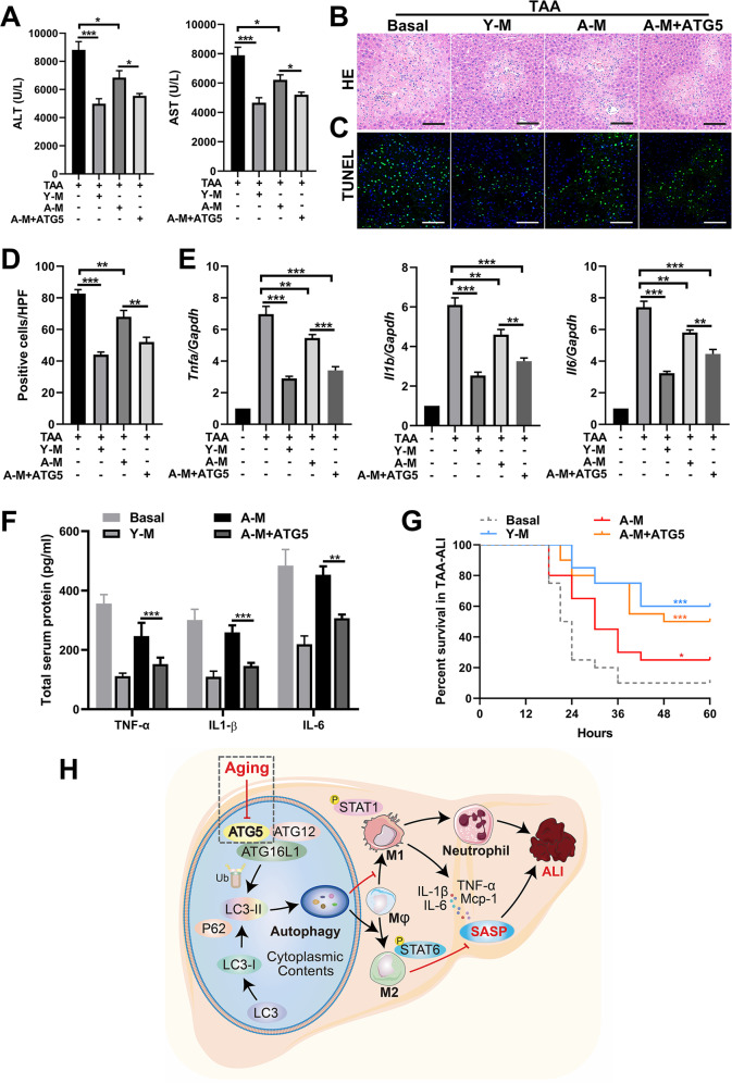
Fig. 8. Administration of BMDMs with ATG5 overexpression relieves TAA-ALI.
Young BMDMs (Y-M) and aged BMDMs (A-M) were isolated and from young and aged mice. Three macrophage populations derived from the mouse BM and the control were prepared: Y-M, A-M, and A-M with LAC-ATG5 (A-M + ATG5). After transplantation of these three kinds of BMDMs by tail vein injection, a TAA-ALI model (200 mg/kg) was established. The results of the transplantation of A-M treated with Torin 1 are shown in S5. A Serum ALT (left panel) and AST (right panel) levels in each group. B Representative H&E staining of the liver. C Representative TUNEL (green fluorescence) staining of the liver with DAPI counterstaining (blue fluorescence) and (D) quantification. E mRNA expression (Tnfa, Il1b, and Il6) in the liver tissue was detected by RT–PCR. The average target gene/GAPDH ratios of different experimental groups relative to the control group are given. F Determination of the total protein content (TNF-α, IL-1β, and IL-6) in the serum by ELISA. Then, a TAA dose of 500 mg/kg was used to calculate mortality. G Summary of mouse mortality in each group. H Model depicting the aggravation of ALI during aging via altered macrophage polarization induced by autophagy deficiency. During aging, macrophage autophagy decreases in mice, which may be ascribed to the downregulation of ATG5. In addition, macrophages are preferentially polarized to the M1 phenotype in ALI, accompanied by increased production of proinflammatory factors (SASP) and massive neutrophil infiltration, thus aggravating ALI. In addition, this aggravation can be reversed by transplantation of macrophages with ATG5 restoration. All data shown represent n = 8–10 mice per group, and 20 mice per group were used for mortality analysis. All results are representative of at least three independent experiments. Values are presented as the mean ± SD. *p ≤ 0.05; **p ≤ 0.01; ***p ≤ 0.001.

