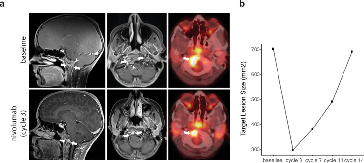
Fig. 3. Response of a poorly differentiated paediatric chordoma to nivolumab.
a Sagittal (left) and axial (middle) post-contrast MRI and concurrent PET/CT (right) images prior to nivolumab therapy (top panel) demonstrate an enhancing solid mass involving the clivus, basiocciput and right occipital condyle (arrows), with effacement of the premedullary cistern and indentation upon the brainstem (arrowheads) and a large focus of metabolic activity at the tumour site (SUVmax 8.2; arrow). Follow-up images (lower panels) 3 months after the initiation of nivolumab therapy show substantial decrease in size of the mass (arrows), with resolution of mass effect upon the brainstem. The concurrent PET/CT image shows attendant decrease in the region of metabolic activity (SUVmax 6.0; arrow), indicating a partial radiographic response. b Target lesion size at baseline and following 3, 7, 11 and 14 cycles of nivolumab.

